Application
Fig1: Western blot analysis of Pax8 on human thyroid gland tissue and SKOV-3 cell lysates using anti-Pax8 antibody at 1/500 dilution.Application
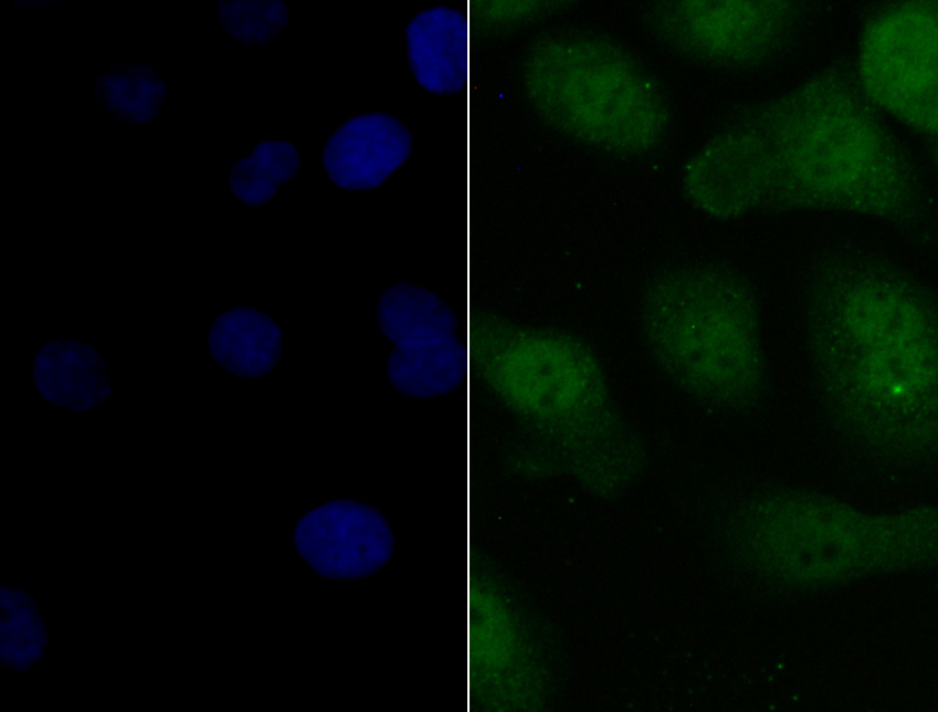
Fig2: ICC staining Pax8 in SKOV-3 cells (green). The nuclear counter stain is DAPI (blue). Cells were fixed in paraformaldehyde, permeabilised with 0.25% Triton X100/PBS.Application
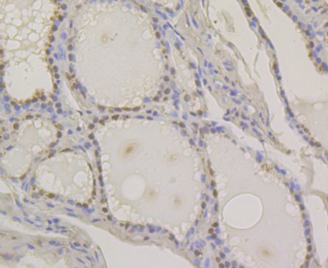
Fig3: Immunohistochemical analysis of paraffin-embedded human thyroid gland tissue using anti-Pax8 antibody. Counter stained with hematoxylin.Application
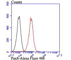
Fig4: Flow cytometric analysis of SiHa cells with Pax8 antibody at 1/100 dilution (red) compared with an unlabelled control (cells without incubation with primary antibody; black). Alexa Fluor 488-conjugated goat anti-rabbit IgG was used as the secondary antibody.| Product Name | Pax8 |
|---|---|
| Antibody Type | Primary Antibodies |
| Antigen Alias | 2-cyclic phosphate 2-phosphohydrolase antibody 35-alpha calcimedin antibody Annexin A3 antibody Annexin III antibody Annexin-3 antibody ANX3 antibody ANXA 3 antibody Anxa3 antibody ANXA3_HUMAN antibody Calcimedin 35 alpha antibody Inositol 1 2 cyclic phosphate 2 phosphohydrolase antibody Inositol 1 antibody Lipocortin III antibody OTTHUMP00000160701 antibody OTTHUMP00000219237 antibody OTTHUMP00000219238 antibody PAP III antibody PAP-III antibody Placental anticoagulant protein III antibody |
| Product description | The Pax family encodes transcription factors that function during embryogenesis and regulate the temporal and position-dependent differentiation of cells. Pax-8 is expressed in the developing and adult thyroid, the developing secretory system and at lower levels, in the adult kidney. Pax-8 complexes with TTF-1 and TTF-2 to induce thyroid follicular cell differentiation and thyroid hormone biosynthesis by regulating the expression of sodium iodide symporter (NIS), thyroid peroxidase (TPO), thyroglobulin (TG) and the thyrotropin receptor (TSHR). Treatment of FRTL-5 cells with TGFβ1 decreases Pax-8 mRNA levels and Pax-8 DNA binding activity, which suppresses the expression of TG and the formation of thyrocytes. Patients who have autosomal dominant mutations of the Pax-8 gene develop thyroid dysgenesis. The Pax-8 gene produces six isoforms, A to F, that are generated by alternative splicing and differ in their carboxy-terminal regions. The Pax-8 isoforms display different DNA binding capacities and are thought to be functionally distinct. The gene which encodes Pax-8 maps to human chromosome 2q12-q14. |
| Immunogen | Synthetic peptide within Human Pax8 aa 400-450. |
| Clonality | Polyclonal |
|---|---|
| Isotype | IgG |
| Host Species | Rabbit |
| Tested Applications | WBICCIHCFC |
| WB:1:500 ICC:1:50-1:200 IHC:1:50-1:200 FC:1:50-1:100 |
|
| Species Reactivity | Human |
| Concentration | 1mg/ml |
| Alternative Names | 2-cyclic phosphate 2-phosphohydrolase antibody 35-alpha calcimedin antibody Annexin A3 antibody Annexin III antibody Annexin-3 antibody ANX3 antibody ANXA 3 antibody Anxa3 antibody ANXA3_HUMAN antibody Calcimedin 35 alpha antibody Inositol 1 2 cyclic phosphate 2 phosphohydrolase antibody Inositol 1 antibody Lipocortin III antibody OTTHUMP00000160701 antibody OTTHUMP00000219237 antibody OTTHUMP00000219238 antibody PAP III antibody PAP-III antibody Placental anticoagulant protein III antibody |
|---|---|
| Molecular Weight(MW) | 48 kDa |
| Cellular Localization | Nucleus. |
| SwissProt ID | Q06710 |
|---|
Application
Fig1: Western blot analysis of Pax8 on human thyroid gland tissue and SKOV-3 cell lysates using anti-Pax8 antibody at 1/500 dilution.
Application
Fig2: ICC staining Pax8 in SKOV-3 cells (green). The nuclear counter stain is DAPI (blue). Cells were fixed in paraformaldehyde, permeabilised with 0.25% Triton X100/PBS.
Application
Fig3: Immunohistochemical analysis of paraffin-embedded human thyroid gland tissue using anti-Pax8 antibody. Counter stained with hematoxylin.
Application
Fig4: Flow cytometric analysis of SiHa cells with Pax8 antibody at 1/100 dilution (red) compared with an unlabelled control (cells without incubation with primary antibody; black). Alexa Fluor 488-conjugated goat anti-rabbit IgG was used as the secondary antibody.| Positive Control | SKOV-3, human thyroid gland tissue, SiHa. |
|---|---|
| Application Notes | WB:1:500 ICC:1:50-1:200 IHC:1:50-1:200 FC:1:50-1:100 |
| Form | Liquid |
|---|---|
| Storage Instructions | Store at +4℃ after thawing. Aliquot store at -20℃. Avoid repeated freeze / thaw cycles. |
| Storage Buffer | 1*TBS (pH7.4), 0.5%BSA, 50%Glycerol. Preservative: 0.05% Sodium Azide. |


