Application
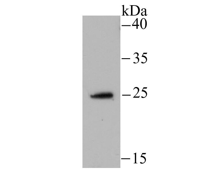
Fig1: Western blot analysis of NDUFS3 on HepG2 cell using anti-NDUFS3 antibody at 1/500 dilution.Application
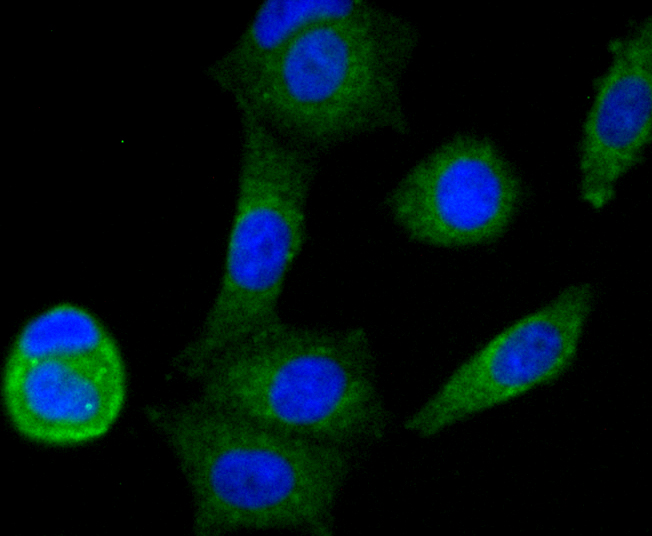
Fig2: ICC staining NDUFS3 in MCF-7 cells (green). The nuclear counter stain is DAPI (blue). Cells were fixed in paraformaldehyde, permeabilised with 0.25% Triton X100/PBS.Application
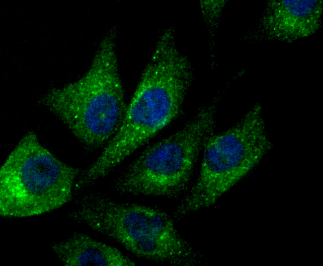
Fig3: ICC staining NDUFS3 in PC-3M cells (green). The nuclear counter stain is DAPI (blue). Cells were fixed in paraformaldehyde, permeabilised with 0.25% Triton X100/PBS.Application
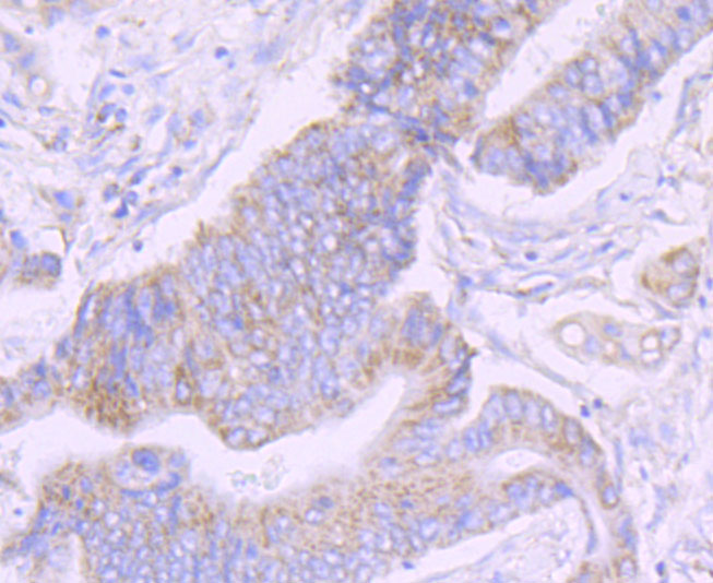
Fig4: Immunohistochemical analysis of paraffin-embedded human colon cancer tissues using anti-NDUFS3 receptor 2 antibody. Counter stained with hematoxylin.Application
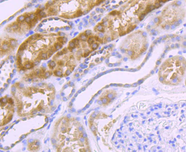
Fig5: Immunohistochemical analysis of paraffin-embedded human kidney cancer tissues using anti-NDUFS3 receptor 2 antibody. Counter stained with hematoxylin.| Product Name | NDUFS3 [JU10-32] |
|---|---|
| Antibody Type | Primary Antibodies |
| Antigen Alias | CI 30 antibody CI 30KD antibody CI-30kD antibody Complex I 30KD antibody Complex I 30kDa subunit antibody COMPLEX I, MITOCHONDRIAL RESPIRATORY CHAIN, 30-KD SUBUNIT antibody Complex I-30kD antibody mitochondrial antibody NADH coenzyme Q reductase antibody NADH dehydrogenase (ubiquinone) Fe S protein 3 30kDa antibody NADH dehydrogenase [ubiquinone] iron sulfur protein 3 mitochondrial antibody NADH dehydrogenase [ubiquinone] iron-sulfur protein 3 antibody NADH dehydrogenase ubiquinone 30 kDa subunit antibody NADH-ubiquinone oxidoreductase 30 kDa subunit antibody NADH-Ubiquinone Oxidoreductase Fe-S Protein 3 antibody NDUFS3 antibody NDUS3_HUMAN antibody |
| Product description | NDUFS3 (NADH dehydrogenase ubiquinone iron-sulfur protein 3) is one of about 45 subunits comprising complex I of the oxidative phosphorylation electron transport chain. The multisubunit NADH: ubiquinone oxidoreductase (complex I) is the first enzyme complex in the electron transport chain of the mitochondria. NDUFS3 is the last subunit of the seven subunits that make up the core of complex I. Through use of chaotropic agents, complex I can be separated into three different fractions: a flavoprotein fraction, an iron-sulfur protein (IP) fraction, and a hydrophobic protein (HP) fraction. The IP fraction includes NDUFS1-7. NDUFS3 contains a highly conserved casein kinase II phosphorylation site. Mutations in the NDUFS3 gene may cause optic atrophy, Leigh syndrome and complex I deficiency. |
| Immunogen | Recombinant protein |
| Clonality | Monoclonal |
|---|---|
| Isotype | IgG |
| Host Species | Recombinant rabbit |
| Tested Applications | WBIHCICCIFIP |
| WB:1:500 IHC:1:50 ICC:1:50 IP:1:10-1:50 |
|
| Species Reactivity | Human |
| Concentration | 1mg/ml |
| Alternative Names | CI 30 antibody
CI 30KD antibody
CI-30kD antibody
Complex I 30KD antibody
Complex I 30kDa subunit antibody
COMPLEX I MITOCHONDRIAL RESPIRATORY CHAIN 30-KD SUBUNIT antibody Complex I-30kD antibody mitochondrial antibody NADH coenzyme Q reductase antibody NADH dehydrogenase (ubiquinone) Fe S protein 3 30kDa antibody NADH dehydrogenase [ubiquinone] iron sulfur protein 3 mitochondrial antibody NADH dehydrogenase [ubiquinone] iron-sulfur protein 3 antibody NADH dehydrogenase ubiquinone 30 kDa subunit antibody NADH-ubiquinone oxidoreductase 30 kDa subunit antibody NADH-Ubiquinone Oxidoreductase Fe-S Protein 3 antibody NDUFS3 antibody NDUS3_HUMAN antibody |
|---|---|
| Molecular Weight(MW) | 25 kDa |
| Cellular Localization | Mitochondrion inner membrane. |
| SwissProt ID | O75489 |
|---|
Application
Fig1: Western blot analysis of NDUFS3 on HepG2 cell using anti-NDUFS3 antibody at 1/500 dilution.
Application
Fig2: ICC staining NDUFS3 in MCF-7 cells (green). The nuclear counter stain is DAPI (blue). Cells were fixed in paraformaldehyde, permeabilised with 0.25% Triton X100/PBS.
Application
Fig3: ICC staining NDUFS3 in PC-3M cells (green). The nuclear counter stain is DAPI (blue). Cells were fixed in paraformaldehyde, permeabilised with 0.25% Triton X100/PBS.
Application
Fig4: Immunohistochemical analysis of paraffin-embedded human colon cancer tissues using anti-NDUFS3 receptor 2 antibody. Counter stained with hematoxylin.
Application
Fig5: Immunohistochemical analysis of paraffin-embedded human kidney cancer tissues using anti-NDUFS3 receptor 2 antibody. Counter stained with hematoxylin.| Positive Control | HepG2, MCF-7, PC-3M, human colon cancer tissues, human kidney tissues. |
|---|---|
| Application Notes | WB:1:500 IHC:1:50 ICC:1:50 IP:1:10-1:50 |
| Form | Liquid |
|---|---|
| Storage Instructions | Store at +4℃ after thawing. Aliquot store at -20℃ or -80℃. Avoid repeated freeze / thaw cycles. |
| Storage Buffer | 1*TBS (pH7.4), 1%BSA, 40%Glycerol. Preservative: 0.05% Sodium Azide. |


