Application
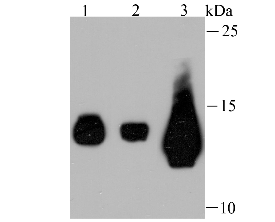
Fig1: Western blot analysis of Fragilis on different cell lysates using anti-Fragilis antibody at 1/500 dilution. Positive control: Lane 1: Hela Lane 2: HepG2 Lane 3: HelaApplication
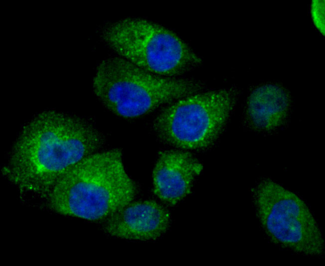
Fig2: ICC staining Fragilis in HUVEC cells (green). The nuclear counter stain is DAPI (blue). Cells were fixed in paraformaldehyde, permeabilised with 0.25% Triton X100/PBS.Application
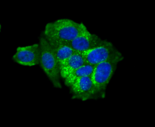
Fig3: ICC staining Fragilis in MCF-7 cells (green). The nuclear counter stain is DAPI (blue). Cells were fixed in paraformaldehyde, permeabilised with 0.25% Triton X100/PBS.Application
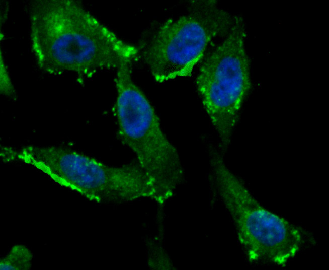
Fig4: ICC staining Fragilis in PC-3M cells (green). The nuclear counter stain is DAPI (blue). Cells were fixed in paraformaldehyde, permeabilised with 0.25% Triton X100/PBS.Application
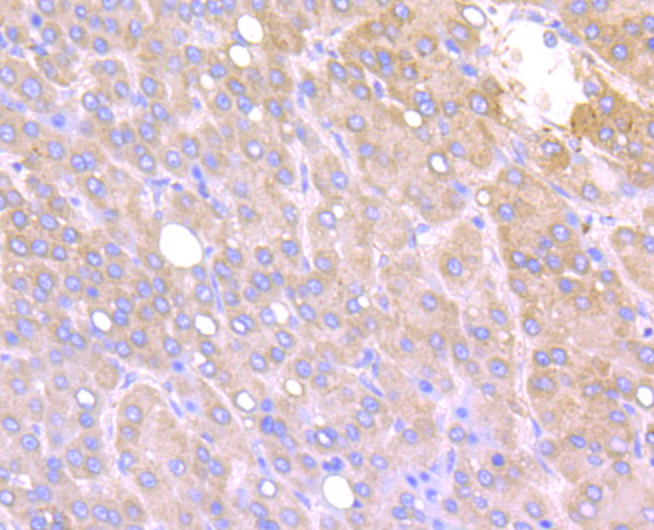
Fig5: Immunohistochemical analysis of paraffin-embedded human liver cancer tissue using anti-Fragilis antibody. Counter stained with hematoxylin.Application
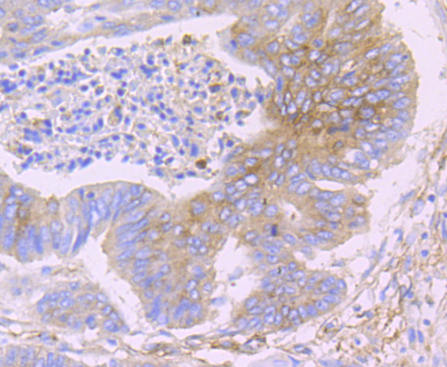
Fig6: Immunohistochemical analysis of paraffin-embedded human colon cancer tissue using anti-Fragilis antibody. Counter stained with hematoxylin.Application
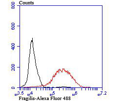
Fig7: Flow cytometric analysis of MCF-7 cells with Fragilis antibody at 1/100 dilution (red) compared with an unlabelled control (cells without incubation with primary antibody; black). Alexa Fluor 488-conjugated goat anti rabbit IgG was used as the secondary antibody.| Product Name | Fragilis [JU73-02] |
|---|---|
| Antibody Type | Primary Antibodies |
| Antigen Alias | 1 8U antibody Fragilis antibody IFITM3 antibody IFM3_HUMAN antibody Interferon induced transmembrane protein 3 (1 8U) antibody Interferon Induced Transmembrane Protein 3 antibody Interferon inducible antibody Interferon inducible protein 1 8U antibody Interferon Inducible Protein 15 antibody Interferon Inducible Protein Homolog antibody Interferon-induced transmembrane protein 3 antibody Interferon-inducible protein 1-8U antibody IP15 antibody |
| Product description | IFN-induced antiviral protein which disrupts intracellular cholesterol homeostasis. Inhibits the entry of viruses to the host cell cytoplasm by preventing viral fusion with cholesterol depleted endosomes. May inactivate new enveloped viruses which buds out of the infected cell, by letting them go out with a cholesterol depleted membrane. Active against multiple viruses, including influenza A virus, SARS coronavirus (SARS-CoV), Marburg virus (MARV) and Ebola virus (EBOV), Dengue virus (DNV), West Nile virus (WNV), human immunodeficiency virus type 1 (HIV-1) and vesicular stomatitis virus (VSV). Can inhibit: influenza virus hemagglutinin protein-mediated viral entry, MARV and EBOV GP1,2-mediated viral entry, SARS-CoV S protein-mediated viral entry and VSV G protein-mediated viral entry. Plays a critical role in the structural stability and function of vacuolar ATPase (v-ATPase). Establishes physical contact with the v-ATPase of endosomes which is critical for proper clathrin localization and is also required for the function of the v-ATPase to lower the pH in phagocytic endosomes thus establishing an antiviral state. |
| Immunogen | Recombinant protein |
| Clonality | Monoclonal |
|---|---|
| Isotype | IgG |
| Host Species | Recombinant rabbit |
| Tested Applications | WBICC/IFIHCFC |
| WB:1:1,000-1:5,000 ICC:1:50-1:200 IHC:1:50-1:100 FC:1:50-1:100 |
|
| Species Reactivity | Human |
| Concentration | 1mg/ml |
| Alternative Names | 1 8U antibody Fragilis antibody IFITM3 antibody IFM3_HUMAN antibody Interferon induced transmembrane protein 3 (1 8U) antibody Interferon Induced Transmembrane Protein 3 antibody Interferon inducible antibody Interferon inducible protein 1 8U antibody Interferon Inducible Protein 15 antibody Interferon Inducible Protein Homolog antibody Interferon-induced transmembrane protein 3 antibody Interferon-inducible protein 1-8U antibody IP15 antibody |
|---|---|
| Molecular Weight(MW) | 15 kDa |
| Cellular Localization | Cell membrane. Lysosome. |
| SwissProt ID | Q01628 |
|---|
Application
Fig1: Western blot analysis of Fragilis on different cell lysates using anti-Fragilis antibody at 1/500 dilution. Positive control: Lane 1: Hela Lane 2: HepG2 Lane 3: Hela
Application
Fig2: ICC staining Fragilis in HUVEC cells (green). The nuclear counter stain is DAPI (blue). Cells were fixed in paraformaldehyde, permeabilised with 0.25% Triton X100/PBS.
Application
Fig3: ICC staining Fragilis in MCF-7 cells (green). The nuclear counter stain is DAPI (blue). Cells were fixed in paraformaldehyde, permeabilised with 0.25% Triton X100/PBS.
Application
Fig4: ICC staining Fragilis in PC-3M cells (green). The nuclear counter stain is DAPI (blue). Cells were fixed in paraformaldehyde, permeabilised with 0.25% Triton X100/PBS.
Application
Fig5: Immunohistochemical analysis of paraffin-embedded human liver cancer tissue using anti-Fragilis antibody. Counter stained with hematoxylin.
Application
Fig6: Immunohistochemical analysis of paraffin-embedded human colon cancer tissue using anti-Fragilis antibody. Counter stained with hematoxylin.
Application
Fig7: Flow cytometric analysis of MCF-7 cells with Fragilis antibody at 1/100 dilution (red) compared with an unlabelled control (cells without incubation with primary antibody; black). Alexa Fluor 488-conjugated goat anti rabbit IgG was used as the secondary antibody.| Positive Control | Hela, HepG2, HUVEC, MCF-7, PC-3M, human liver cancer tissue, human colon cancer tissue. |
|---|---|
| Application Notes | WB:1:1,000-1:5,000 ICC:1:50-1:200 IHC:1:50-1:100 FC:1:50-1:100 |
| Form | Liquid |
|---|---|
| Storage Instructions | Store at +4℃ after thawing. Aliquot store at -20℃ or -80℃. Avoid repeated freeze / thaw cycles. |
| Storage Buffer | 1*TBS (pH7.4), 1%BSA, 40%Glycerol. Preservative: 0.05% Sodium Azide. |


