Application
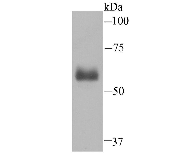
Fig1: Western blot analysis of Hemopexin on human plasma lysate using anti-Hemopexin antibody at 1/500 dilution.Application
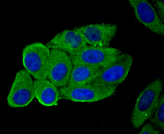
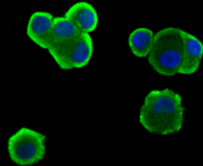
Fig2: ICC staining Hemopexin in HepG2 cells (green). The nuclear counter stain is DAPI (blue). Cells were fixed in paraformaldehyde, permeabilised with 0.25% Triton X100/PBS.Application
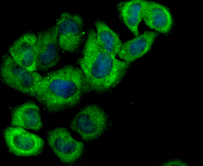
Fig3: ICC staining Hemopexin in LO2 cells (green). The nuclear counter stain is DAPI (blue). Cells were fixed in paraformaldehyde, permeabilised with 0.25% Triton X100/PBS.Application
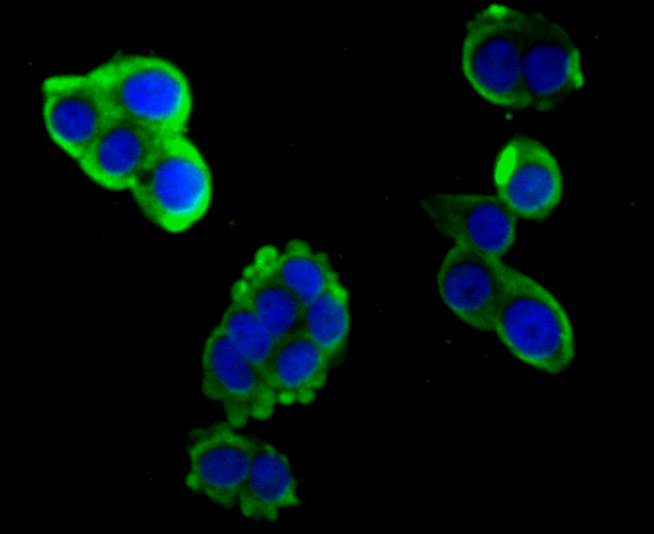
Fig4: ICC staining Hemopexin in LOVO cells (green). The nuclear counter stain is DAPI (blue). Cells were fixed in paraformaldehyde, permeabilised with 0.25% Triton X100/PBS.Application
Fig5: ICC staining Hemopexin in SK-Br-3 cells (green). The nuclear counter stain is DAPI (blue). Cells were fixed in paraformaldehyde, permeabilised with 0.25% Triton X100/PBS.Application
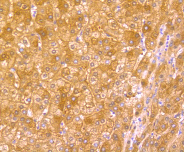
Fig6: Immunohistochemical analysis of paraffin-embedded human liver tissue using anti-Hemopexin antibody. Counter stained with hematoxylin.Application
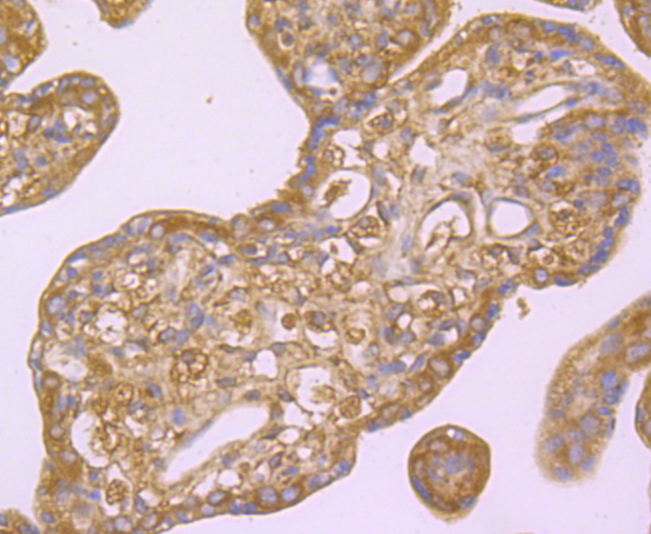
Fig7: Immunohistochemical analysis of paraffin-embedded human placenta tissue using anti-Hemopexin antibody. Counter stained with hematoxylin.| Product Name | Hemopexin [JU83-33] |
|---|---|
| Antibody Type | Primary Antibodies |
| Antigen Alias | Beta 1B glycoprotein antibody Beta-1B-glycoprotein antibody FLJ56652 antibody HEMO antibody HEMO_HUMAN antibody Hemopexin antibody Hpx antibody HX antibody |
| Product description | Hemopexin (also known as β 1B glycoprotein or HPX), a 462 amino acid protein, functions as a scavenger and transporter of toxic plasma heme, transporting heme to the liver for breakdown and iron recovery. Hemopexin cooperates with Albumin, Haptoglobin, and high and low density lipoproteins to trap toxic plasma heme, which occurs as the result of the degradation of hemoglobin, myoglobin and enzymes with heme prosthetic groups, and to ensure the clearance of toxic heme from the plasma. After releasing the heme molecule, the free Hemopexin returns to circulation. It is expressed by the liver and is secreted in plasma. Hemopexin may play a role in the maintenance of metal ion homeostasis. It binds the following metal ions in order of highest to lowest affinity: nickel, copper, cobalt, zinc and manganese. Hemopexin can also act as a toxic protease that leads to proteinuria and glomerular alterations, which are characteristics of minimal changes disease (MCD), a common cause of nephrotic syndrome. |
| Immunogen | Recombinant protein |
| Clonality | Monoclonal |
|---|---|
| Isotype | IgG |
| Host Species | Recombinant rabbit |
| Tested Applications | WBICC/IFIHCIP |
| WB:1:500-1:2,000 ICC:1:50-1:200 IHC:1:100-1:500 IP:1:10-1:50 |
|
| Species Reactivity | Human |
| Concentration | 1mg/ml |
| Alternative Names | Beta 1B glycoprotein antibody Beta-1B-glycoprotein antibody FLJ56652 antibody HEMO antibody HEMO_HUMAN antibody Hemopexin antibody Hpx antibody HX antibody |
|---|---|
| Molecular Weight(MW) | 52 kDa |
| Cellular Localization | Secreted. |
| SwissProt ID | P02790 |
|---|
Application
Fig1: Western blot analysis of Hemopexin on human plasma lysate using anti-Hemopexin antibody at 1/500 dilution.
Application
Fig2: ICC staining Hemopexin in HepG2 cells (green). The nuclear counter stain is DAPI (blue). Cells were fixed in paraformaldehyde, permeabilised with 0.25% Triton X100/PBS.
Application
Fig3: ICC staining Hemopexin in LO2 cells (green). The nuclear counter stain is DAPI (blue). Cells were fixed in paraformaldehyde, permeabilised with 0.25% Triton X100/PBS.
Application
Fig4: ICC staining Hemopexin in LOVO cells (green). The nuclear counter stain is DAPI (blue). Cells were fixed in paraformaldehyde, permeabilised with 0.25% Triton X100/PBS.
Application
Fig5: ICC staining Hemopexin in SK-Br-3 cells (green). The nuclear counter stain is DAPI (blue). Cells were fixed in paraformaldehyde, permeabilised with 0.25% Triton X100/PBS.
Application
Fig6: Immunohistochemical analysis of paraffin-embedded human liver tissue using anti-Hemopexin antibody. Counter stained with hematoxylin.
Application
Fig7: Immunohistochemical analysis of paraffin-embedded human placenta tissue using anti-Hemopexin antibody. Counter stained with hematoxylin.| Positive Control | Human plasma lysate, HepG2, LO2, LOVO, SK-Br-3, human liver tissue, human placenta tissue. |
|---|---|
| Application Notes | WB:1:500-1:2,000 ICC:1:50-1:200 IHC:1:100-1:500 IP:1:10-1:50 |
| Form | Liquid |
|---|---|
| Storage Instructions | Store at +4℃ after thawing. Aliquot store at -20℃ or -80℃. Avoid repeated freeze / thaw cycles. |
| Storage Buffer | 1*TBS (pH7.4), 1%BSA, 40%Glycerol. Preservative: 0.05% Sodium Azide. |


