Application
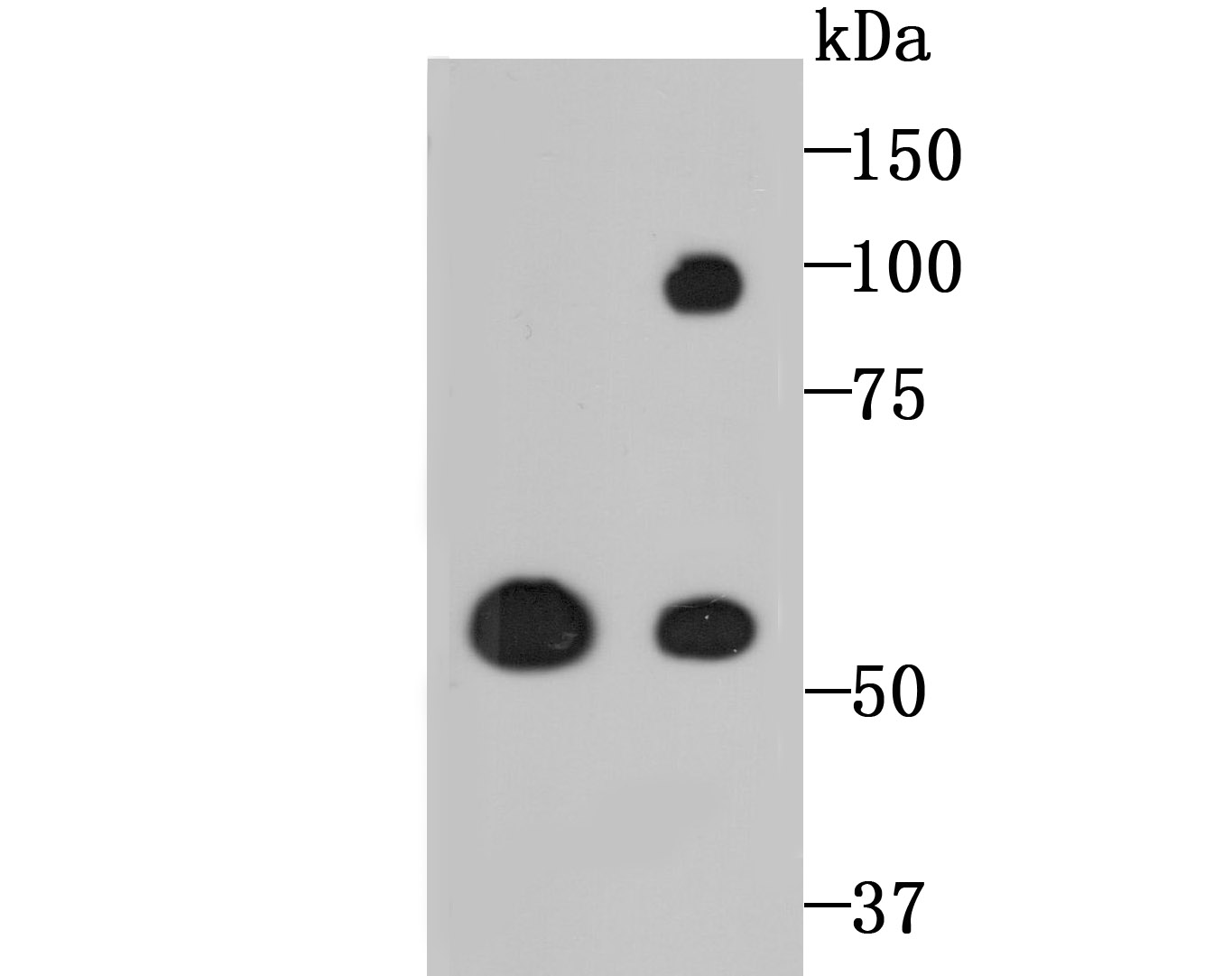
Fig1: Western blot analysis of LOXL2 on PC-12 and A549 cell lysates using anti-LOXL2 antibody at 1/500 dilution.Application
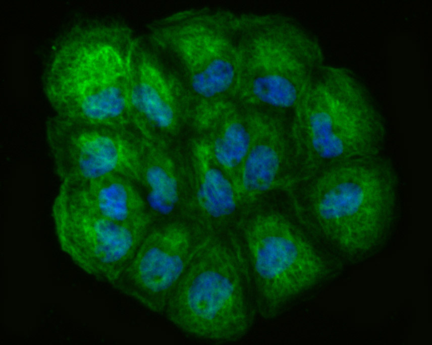
Fig2: ICC staining LOXL2 in A431 cells (green). The nuclear counter stain is DAPI (blue). Cells were fixed in paraformaldehyde, permeabilised with 0.25% Triton X100/PBS.Application
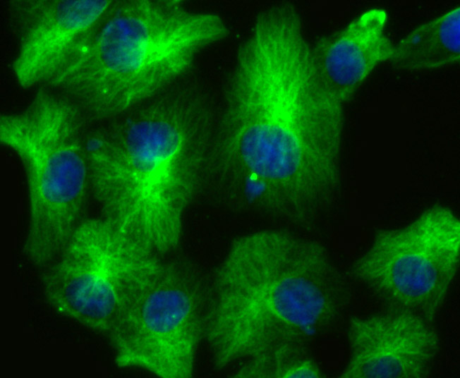
Fig3: ICC staining LOXL2 in A549 cells (green). The nuclear counter stain is DAPI (blue). Cells were fixed in paraformaldehyde, permeabilised with 0.25% Triton X100/PBS.Application
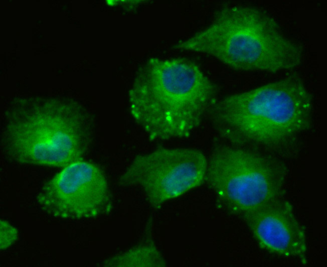
Fig4: ICC staining LOXL2 in HUVEC cells (green). The nuclear counter stain is DAPI (blue). Cells were fixed in paraformaldehyde, permeabilised with 0.25% Triton X100/PBS.Application
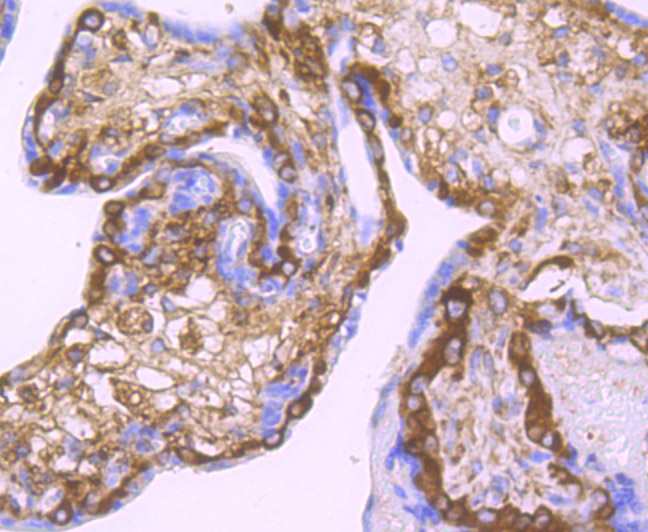
Fig5: Immunohistochemical analysis of paraffin-embedded human placenta tissue using anti-LOXL2 antibody. Counter stained with hematoxylin.Application
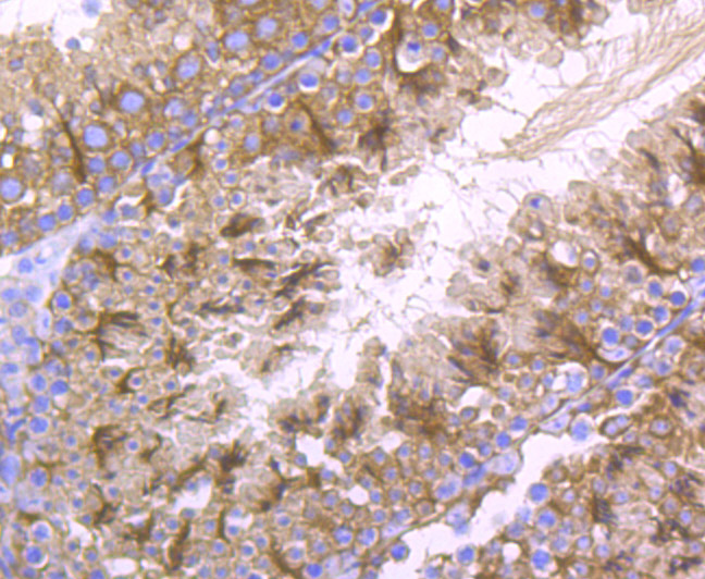
Fig6: Immunohistochemical analysis of paraffin-embedded mouse testis tissue using anti-LOXL2 antibody. Counter stained with hematoxylin.Application
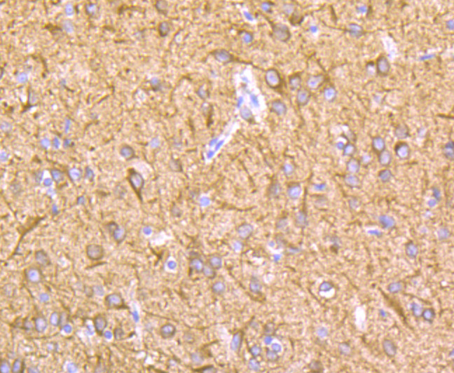
Fig7: Immunohistochemical analysis of paraffin-embedded rat brain tissue using anti-LOXL2 antibody. Counter stained with hematoxylin.Application
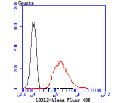
Fig8: Flow cytometric analysis of A549 cells with LOXL2 antibody at 1/100 dilution (red) compared with an unlabelled control (cells without incubation with primary antibody; black). Alexa Fluor 488-conjugated goat anti rabbit IgG was used as the secondary antibody.| Product Name | LOXL2 [JU32-53] |
|---|---|
| Antibody Type | Primary Antibodies |
| Antigen Alias | LOR 2 antibody LOR2 antibody LOX L2 antibody LOXL 2 antibody LOXL2 antibody LOXL2_HUMAN antibody Lysyl oxidase homolog 2 antibody Lysyl oxidase like 2 antibody Lysyl oxidase like protein 2 antibody Lysyl oxidase related 2 antibody Lysyl oxidase related protein 2 antibody Lysyl oxidase related protein WS9 14 antibody Lysyl oxidase-like protein 2 antibody Lysyl oxidase-related protein 2 antibody Lysyl oxidase-related protein WS9-14 antibody WS9 14 antibody |
| Product description | Mediates the post-translational oxidative deamination of lysine residues on target proteins leading to the formation of deaminated lysine (allysine). When secreted in extracellular matrix, promotes cross-linking of extracellular matrix proteins by mediating oxidative deamination of peptidyl lysine residues in precursors to fibrous collagen and elastin. Acts as a regulator of sprouting angiogenesis, probably via collagen IV scaffolding. When nuclear, acts as a transcription corepressor and specifically mediates deamination of trimethylated 'Lys-4' of histone H3 (H3K4me3), a specific tag for epigenetic transcriptional activation. Involved in epithelial to mesenchymal transition (EMT) via interaction with SNAI1 and participates in repression of E-cadherin, probably by mediating deamination of histone H3. Also involved in E-cadherin repression following hypoxia, a hallmark of epithelial to mesenchymal transition believed to amplify tumor aggressiveness, suggesting that it may play a role in tumor progression. Acts as a regulator of chondrocyte differentiation, probably by regulating expression of factors that control chondrocyte differentiation. |
| Immunogen | Recombinant protein |
| Clonality | Monoclonal |
|---|---|
| Isotype | IgG |
| Host Species | Recombinant rabbit |
| Tested Applications | WBICCIFIHCFC |
| WB:1:500-1:2,000 ICC:1:50-1:200 IHC:1:50-1:100 FC:1:50-1:100 |
|
| Species Reactivity | HumanMouseRat |
| Concentration | 1mg/ml |
| Alternative Names | LOR 2 antibody LOR2 antibody LOX L2 antibody LOXL 2 antibody LOXL2 antibody LOXL2_HUMAN antibody Lysyl oxidase homolog 2 antibody Lysyl oxidase like 2 antibody Lysyl oxidase like protein 2 antibody Lysyl oxidase related 2 antibody Lysyl oxidase related protein 2 antibody Lysyl oxidase related protein WS9 14 antibody Lysyl oxidase-like protein 2 antibody Lysyl oxidase-related protein 2 antibody Lysyl oxidase-related protein WS9-14 antibody WS9 14 antibody |
|---|---|
| Molecular Weight(MW) | 87 kDa, additional band 55 kDa |
| Cellular Localization | Secreted, Nucleus. |
Application
Fig1: Western blot analysis of LOXL2 on PC-12 and A549 cell lysates using anti-LOXL2 antibody at 1/500 dilution.
Application
Fig2: ICC staining LOXL2 in A431 cells (green). The nuclear counter stain is DAPI (blue). Cells were fixed in paraformaldehyde, permeabilised with 0.25% Triton X100/PBS.
Application
Fig3: ICC staining LOXL2 in A549 cells (green). The nuclear counter stain is DAPI (blue). Cells were fixed in paraformaldehyde, permeabilised with 0.25% Triton X100/PBS.
Application
Fig4: ICC staining LOXL2 in HUVEC cells (green). The nuclear counter stain is DAPI (blue). Cells were fixed in paraformaldehyde, permeabilised with 0.25% Triton X100/PBS.
Application
Fig5: Immunohistochemical analysis of paraffin-embedded human placenta tissue using anti-LOXL2 antibody. Counter stained with hematoxylin.
Application
Fig6: Immunohistochemical analysis of paraffin-embedded mouse testis tissue using anti-LOXL2 antibody. Counter stained with hematoxylin.
Application
Fig7: Immunohistochemical analysis of paraffin-embedded rat brain tissue using anti-LOXL2 antibody. Counter stained with hematoxylin.
Application
Fig8: Flow cytometric analysis of A549 cells with LOXL2 antibody at 1/100 dilution (red) compared with an unlabelled control (cells without incubation with primary antibody; black). Alexa Fluor 488-conjugated goat anti rabbit IgG was used as the secondary antibody.| Positive Control | PC-12, A431, A549, HUVEC, human placenta tissue, mouse testistissue, rat brain tissue. |
|---|---|
| Application Notes | WB:1:500-1:2,000 ICC:1:50-1:200 IHC:1:50-1:100 FC:1:50-1:100 |
| Form | Liquid |
|---|---|
| Storage Instructions | Store at +4℃ after thawing. Aliquot store at -20℃ or -80℃. Avoid repeated freeze / thaw cycles. |
| Storage Buffer | 1*TBS (pH7.4), 1%BSA, 40%Glycerol. Preservative: 0.05% Sodium Azide. |


