Application
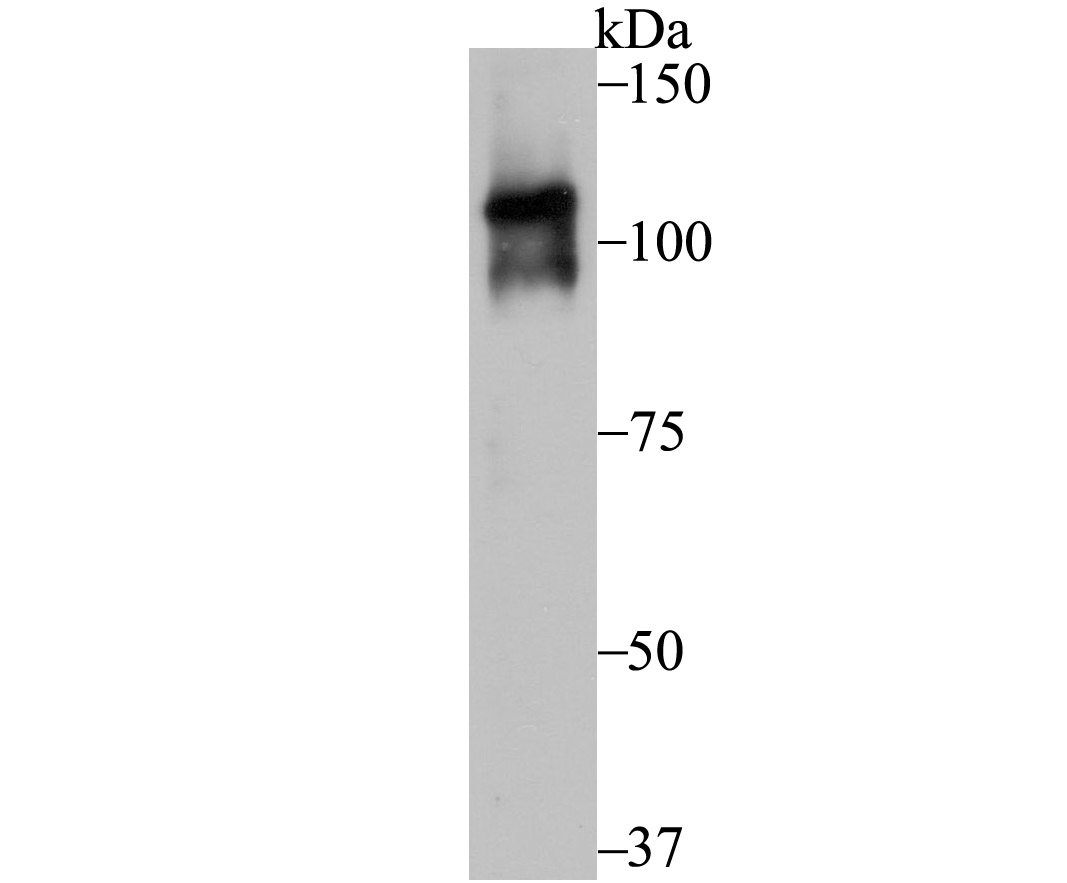
Fig1: Western blot analysis of Dnmt3b on K562 cell lysate using anti-Dnmt3b antibody at 1/500 dilution.Application
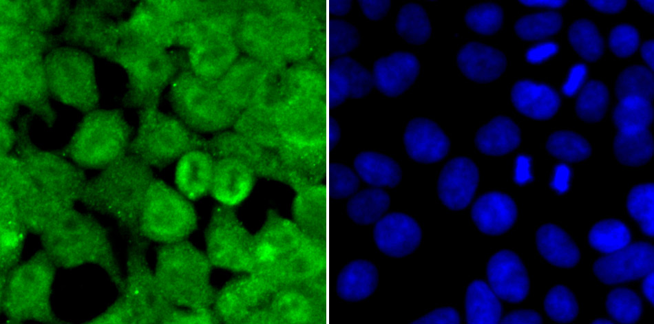
Fig2: ICC staining Dnmt3b in 293T cells (green). The nuclear counter stain is DAPI (blue). Cells were fixed in paraformaldehyde, permeabilised with 0.25% Triton X100/PBS.Application
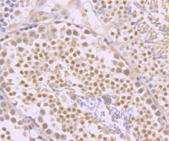
Fig3: Immunohistochemical analysis of paraffin-embedded mouse testis tissue using anti-Dnmt3b antibody. Counter stained with hematoxylin.Application
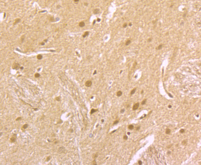
Fig4: Immunohistochemical analysis of paraffin-embedded rat brain tissue using anti-Dnmt3b antibody. Counter stained with hematoxylin.Application
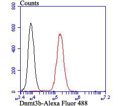
Fig5: Flow cytometric analysis of K562 cells with Dnmt3b antibody at 1/100 dilution (red) compared with an unlabelled control (cells without incubation with primary antibody; black). Alexa Fluor 488-conjugated goat anti-rabbit IgG was used as the secondary antibody.| Product Name | Dnmt3b |
|---|---|
| Antibody Type | Primary Antibodies |
| Antigen Alias | Cytosine 5methyltransferase 3B antibody DNA antibody DNA (cytosine 5) methyltransferase 3 beta antibody DNA (cytosine 5)-methyltransferase 3B antibody DNA (cytosine-5)-methyltransferase 3B antibody DNA methyltransferase HsaIIIB antibody DNA MTase HsaIIIB antibody DNM3B_HUMAN antibody Dnmt3b antibody EC 2.1.1.37 antibody ICF antibody ICF1 antibody M.HsaIIIB antibody MGC124407 antibody RP23-89H14.3 antibody |
| Product description | Methylation at the 5'-position of cytosine is the only known naturally occurring covalent modification of the mammalian genome. DNA methylation requires the enzymatic activity of DNA 5-cytosine methyltransferase (Dnmt) proteins, which catalyze the transfer of a methyl group from S-adenosyl methionine to the 5'-position of cytosines residing in the dinucleotide CpG motif, and this methylation results in transcriptional repression of the target gene. The Dnmt enzymes are encoded by independent genes. Dnmt1 is the most abundant, and it preferentially methylates hemimethylated DNA and coordinates gene expression during development. Additional mammalian Dnmt proteins include Dnmt2 and Dnmt3. Dnmt2 lacks the large N-terminal regulator domain of Dnmt1, is expressed at substantially lower levels in adult tissues, and is likely involved in methylating newly integrated retroviral DNA. Dnmt3a and Dnmt3b are encoded by two distinct genes, but both are abundantly expressed in embryonic stem cells, where they also methylate CpG motifs on DNA. |
| Immunogen | Synthetic peptide within human Dnmt3b aa 1-50. |
| Clonality | Polyclonal |
|---|---|
| Isotype | IgG |
| Host Species | Rabbit |
| Tested Applications | WBICCIHCFC |
| WB:1:500-1:1,000 ICC:1:50-1:200 IHC:1:50-1:200 FC:1:50-1:100 |
|
| Species Reactivity | HumanMouseRat |
| Concentration | 1mg/ml |
| Alternative Names | Cytosine 5methyltransferase 3B antibody DNA antibody DNA (cytosine 5) methyltransferase 3 beta antibody DNA (cytosine 5)-methyltransferase 3B antibody DNA (cytosine-5)-methyltransferase 3B antibody DNA methyltransferase HsaIIIB antibody DNA MTase HsaIIIB antibody DNM3B_HUMAN antibody Dnmt3b antibody EC 2.1.1.37 antibody ICF antibody ICF1 antibody M.HsaIIIB antibody MGC124407 antibody RP23-89H14.3 antibody |
|---|---|
| Molecular Weight(MW) | 96 kDa |
| Cellular Localization | Nucleus. |
Application
Fig1: Western blot analysis of Dnmt3b on K562 cell lysate using anti-Dnmt3b antibody at 1/500 dilution.
Application
Fig2: ICC staining Dnmt3b in 293T cells (green). The nuclear counter stain is DAPI (blue). Cells were fixed in paraformaldehyde, permeabilised with 0.25% Triton X100/PBS.
Application
Fig3: Immunohistochemical analysis of paraffin-embedded mouse testis tissue using anti-Dnmt3b antibody. Counter stained with hematoxylin.
Application
Fig4: Immunohistochemical analysis of paraffin-embedded rat brain tissue using anti-Dnmt3b antibody. Counter stained with hematoxylin.
Application
Fig5: Flow cytometric analysis of K562 cells with Dnmt3b antibody at 1/100 dilution (red) compared with an unlabelled control (cells without incubation with primary antibody; black). Alexa Fluor 488-conjugated goat anti-rabbit IgG was used as the secondary antibody.| Positive Control | K562, 293T, mouse testis tissue, rat brain tissue. |
|---|---|
| Application Notes | WB:1:500-1:1,000 ICC:1:50-1:200 IHC:1:50-1:200 FC:1:50-1:100 |
| Form | Liquid |
|---|---|
| Storage Instructions | Store at +4℃ after thawing. Aliquot store at -20℃. Avoid repeated freeze / thaw cycles. |
| Storage Buffer | 1*TBS (pH7.4), 0.5%BSA, 50%Glycerol. Preservative: 0.05% Sodium Azide. |


