Application
Fig1: Western blot analysis of B7-H4(VTCN1) on SKBR3 cell and human colon tissue lysates using anti-B7-H4(VTCN1) antibody at 1/500 dilution.Application
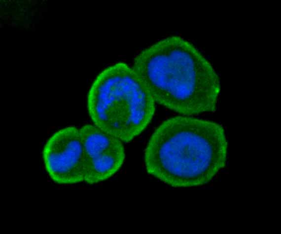
Fig2: ICC staining B7-H4(VTCN1) in MCF-7 cells (green). The nuclear counter stain is DAPI (blue). Cells were fixed in paraformaldehyde, permeabilised with 0.25% Triton X100/PBS.Application
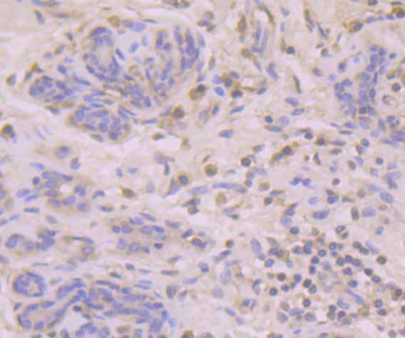
Fig3: Immunohistochemical analysis of paraffin-embedded human breast tissue using anti-B7-H4(VTCN1) antibody. Counter stained with hematoxylin.Application
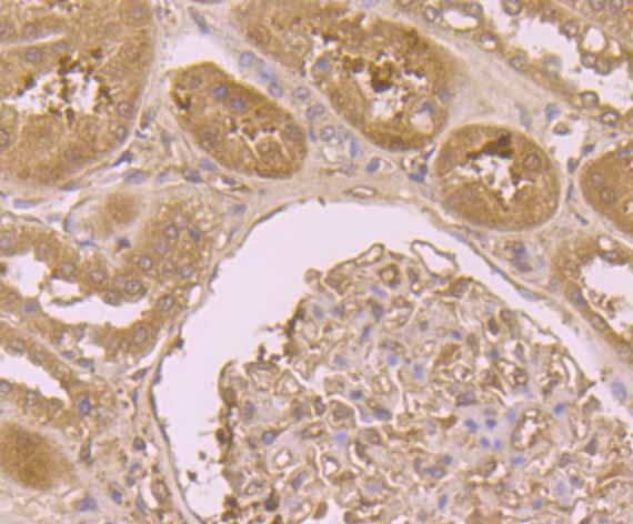
Fig4: Immunohistochemical analysis of paraffin-embedded human kidney tissue using anti-B7-H4(VTCN1) antibody. Counter stained with hematoxylin.Application
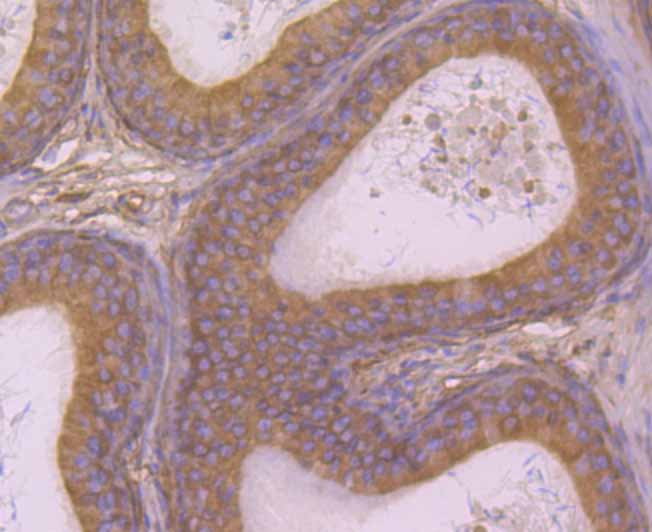
Fig5: Immunohistochemical analysis of paraffin-embedded rat epididymis tissue using anti-B7-H4(VTCN1) antibody. Counter stained with hematoxylin.Application
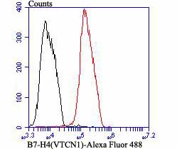
Fig6: Flow cytometric analysis of MCF-7 cells with B7-H4(VTCN1) antibody at 1/100 dilution (red) compared with an unlabelled control (cells without incubation with primary antibody; black). Alexa Fluor 488-conjugated goat anti-rabbit IgG was used as the secondary antibody.| Product Name | B7-H4(VTCN1) |
|---|---|
| Antibody Type | Primary Antibodies |
| Antigen Alias | B7 family member, H4 antibody B7 H4 antibody B7 homolog 4 antibody B7 superfamily member 1 antibody B7 superfamily, member 1 antibody B7-H4 antibody B7h.5 antibody B7h4 antibody B7S1 antibody B7x antibody BC032925 antibody Immune costimulatory protein B7-H4 antibody Immune costimulatory protein B7H4 antibody MGC41287 antibody PRO1291 antibody Protein B7S1 antibody RP11 229A19.4 antibody T cell costimulatory molecule B7x antibody T-cell costimulatory molecule B7x antibody V set domain-containing T cell activation inhibitor 1 antibody V-set domain-containing T-cell activation inhibitor 1 antibody VCTN1 antibody Vtcn1 antibody VTCN1_HUMAN antibody |
| Product description | T cell activation and immune function are regulated by the innate immune system through positive and negative costimulatory proteins. One such protein, B7-H4 (B7-homolog 4, also designated VTCN1), belongs to the B7 Immunoglobulin superfamily of ligand-lymphocyte interacting proteins. Expressed primarily on the membrane of lymphoid cells, B7-H4 is an immunoinhibitory protein that interacts with receptors on the surface of T lymphocytes, thus mediating cellular and humoral immune responses. Overexpression of the B7-H4 protein is associated with certain malignancies, including ovarian and breast cancer, as its interaction with T cells supresses tumor-associated immunity. Current research suggests that, similar to Mucin 16 (CA125), B7-H4 may be a useful biomarker for the early detection of ovarian cancer. |
| Immunogen | Recombinant protein with human B7-H4 aa 50-220. |
| Clonality | Polyclonal |
|---|---|
| Isotype | IgG |
| Host Species | Rabbit |
| Tested Applications | WBICCIHCFC |
| WB:1:500-1:2000 ICC:1:1,000 IHC:1:50-1:200 FC:1:50-1:100 |
|
| Species Reactivity | HumanRat |
| Concentration | 1mg/ml |
| Alternative Names | B7 family member H4 antibody B7 H4 antibody B7 homolog 4 antibody B7 superfamily member 1 antibody B7 superfamily member 1 antibody B7-H4 antibody B7h.5 antibody B7h4 antibody B7S1 antibody B7x antibody BC032925 antibody Immune costimulatory protein B7-H4 antibody Immune costimulatory protein B7H4 antibody MGC41287 antibody PRO1291 antibody Protein B7S1 antibody RP11 229A19.4 antibody T cell costimulatory molecule B7x antibody T-cell costimulatory molecule B7x antibody V set domain-containing T cell activation inhibitor 1 antibody V-set domain-containing T-cell activation inhibitor 1 antibody VCTN1 antibody Vtcn1 antibody VTCN1_HUMAN antibody |
|---|---|
| Molecular Weight(MW) | 31/21/10 kDa (Predicted band size) |
| Cellular Localization | Cell membrane. |
Application
Fig1: Western blot analysis of B7-H4(VTCN1) on SKBR3 cell and human colon tissue lysates using anti-B7-H4(VTCN1) antibody at 1/500 dilution.
Application
Fig2: ICC staining B7-H4(VTCN1) in MCF-7 cells (green). The nuclear counter stain is DAPI (blue). Cells were fixed in paraformaldehyde, permeabilised with 0.25% Triton X100/PBS.
Application
Fig3: Immunohistochemical analysis of paraffin-embedded human breast tissue using anti-B7-H4(VTCN1) antibody. Counter stained with hematoxylin.
Application
Fig4: Immunohistochemical analysis of paraffin-embedded human kidney tissue using anti-B7-H4(VTCN1) antibody. Counter stained with hematoxylin.
Application
Fig5: Immunohistochemical analysis of paraffin-embedded rat epididymis tissue using anti-B7-H4(VTCN1) antibody. Counter stained with hematoxylin.
Application
Fig6: Flow cytometric analysis of MCF-7 cells with B7-H4(VTCN1) antibody at 1/100 dilution (red) compared with an unlabelled control (cells without incubation with primary antibody; black). Alexa Fluor 488-conjugated goat anti-rabbit IgG was used as the secondary antibody.| Positive Control | SKBR3 cell and human colon tissue lysates, MCF-7, rat epididymid tissue, human breast tissue, human breast tissue, human kidney tissue. |
|---|---|
| Application Notes | WB:1:500-1:2000 ICC:1:1,000 IHC:1:50-1:200 FC:1:50-1:100 |
| Form | Liquid |
|---|---|
| Storage Instructions | Store at +4℃ after thawing. Aliquot store at -20℃. Avoid repeated freeze / thaw cycles. |
| Storage Buffer | 1*TBS (pH7.4), 0.5%BSA, 50%Glycerol. Preservative: 0.05% Sodium Azide. |


