Application
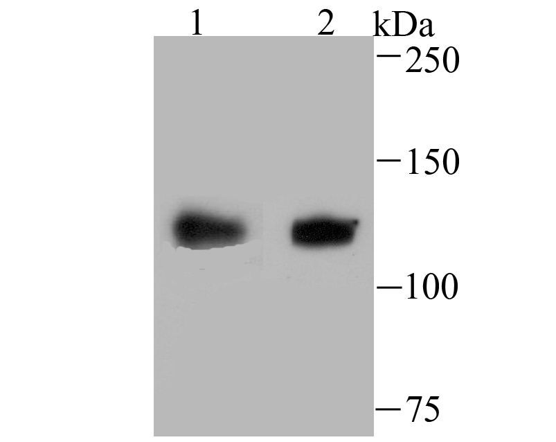
Fig1: Western blot analysis of USP10 on SiHa (1) and A549 (2) cell lysate using anti-USP10 antibody at 1/500 dilution.Application
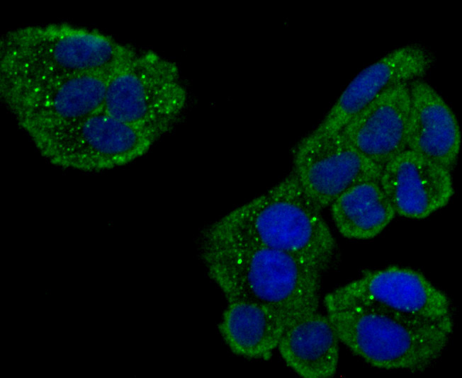
Fig2: ICC staining USP10 in Hela cells (green). The nuclear counter stain is DAPI (blue). Cells were fixed in paraformaldehyde, permeabilised with 0.25% Triton X100/PBS.Application
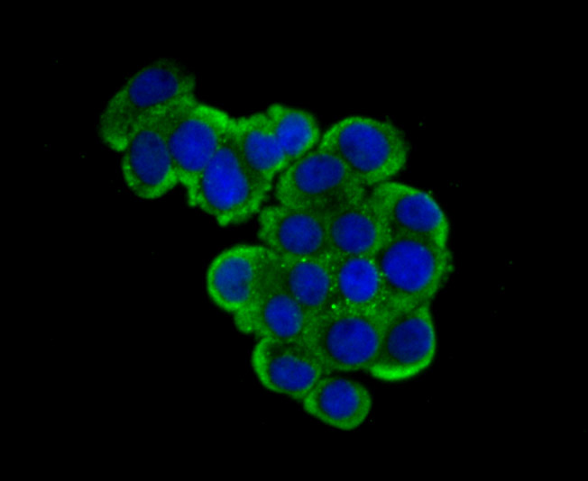
Fig3: ICC staining USP10 in LOVO cells (green). The nuclear counter stain is DAPI (blue). Cells were fixed in paraformaldehyde, permeabilised with 0.25% Triton X100/PBS.Application
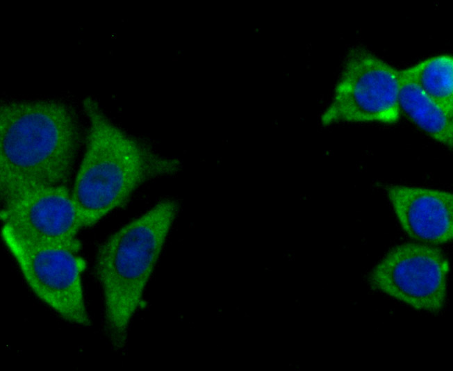
Fig4: ICC staining USP10 in MCF-7 cells (green). The nuclear counter stain is DAPI (blue). Cells were fixed in paraformaldehyde, permeabilised with 0.25% Triton X100/PBS.Application
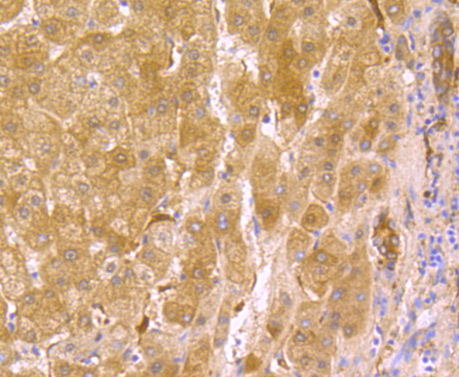
Fig5: Immunohistochemical analysis of paraffin-embedded human liver tissue using anti-USP10 antibody. Counter stained with hematoxylin.Application
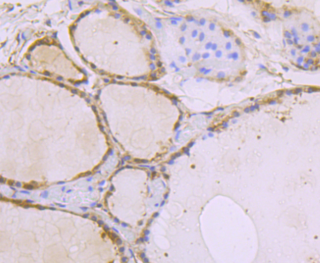
Fig6: Immunohistochemical analysis of paraffin-embedded human thyroid grand tissue using anti-USP10 antibody. Counter stained with hematoxylin.Application
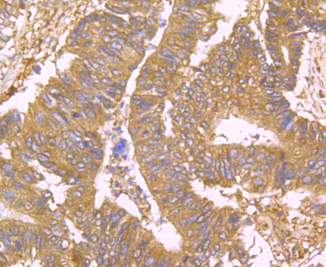
Fig7: Immunohistochemical analysis of paraffin-embedded human colon cancer tissue using anti-USP10 antibody. Counter stained with hematoxylin.Application
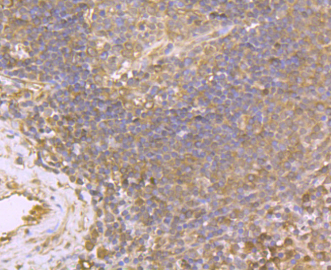
Fig8: Immunohistochemical analysis of paraffin-embedded human spleen tissue using anti-USP10 antibody. Counter stained with hematoxylin.Application
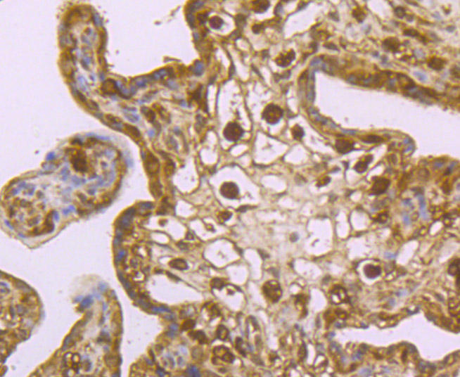
Fig9: Immunohistochemical analysis of paraffin-embedded human placenta tissue using anti-USP10 antibody. Counter stained with hematoxylin.| Product Name | USP10 [JU32-62] |
|---|---|
| Antibody Type | Primary Antibodies |
| Antigen Alias | Deubiquitinating enzyme 10 antibody KIAA0190 antibody MGC124997 antibody MGC2621 antibody Ubiquitin carboxyl terminal hydrolase 10 antibody Ubiquitin carboxyl-terminal hydrolase 10 antibody Ubiquitin specific peptidase 10 antibody Ubiquitin specific processing protease 10 antibody Ubiquitin specific protease 10 antibody Ubiquitin thioesterase 10 antibody Ubiquitin thiolesterase 10 antibody Ubiquitin-specific-processing protease 10 antibody UBP10_HUMAN antibody UBPO antibody USP 10 antibody USP10 antibody |
| Product description | The ubiquitin (Ub) pathway involves three sequential enzymatic steps that facilitate the conjugation of Ub and Ub-like molecules to specific protein substrates. Through the use of a wide range of enzymes that can add or remove ubiquitin, the Ub pathway controls many intracellular processes such as signal transduction, transcriptional activation and cell cycle progression. USP10 (ubiquitin specific peptidase 10), also known as UBPO, is a 798 amino acid protein that belongs to the ubiquitin-specific protease family of cysteine proteases. Expressed in a variety of tissues, USP10 functions to catalyze the cleavage of ubiquitin from ubiquitin-conjugated protein substrates, possibly playing a role in the activity of the DNA-bound androgen receptor complex. |
| Immunogen | Recombinant protein |
| Clonality | Monoclonal |
|---|---|
| Isotype | IgG |
| Host Species | Recombinant rabbit |
| Tested Applications | WBICC/IFIHCIP |
| WB:1:500-1:2,000 ICC:1:50-1:200 IHC:1:50-1:200 |
|
| Species Reactivity | Human |
| Concentration | 1mg/ml |
| Alternative Names | Deubiquitinating enzyme 10 antibody KIAA0190 antibody MGC124997 antibody MGC2621 antibody Ubiquitin carboxyl terminal hydrolase 10 antibody Ubiquitin carboxyl-terminal hydrolase 10 antibody Ubiquitin specific peptidase 10 antibody Ubiquitin specific processing protease 10 antibody Ubiquitin specific protease 10 antibody Ubiquitin thioesterase 10 antibody Ubiquitin thiolesterase 10 antibody Ubiquitin-specific-processing protease 10 antibody UBP10_HUMAN antibody UBPO antibody USP 10 antibody USP10 antibody |
|---|---|
| Molecular Weight(MW) | 110 kDa |
| Cellular Localization | Cytoplasm. Nucleus. |
| SwissProt ID | Q14694 |
|---|
Application
Fig1: Western blot analysis of USP10 on SiHa (1) and A549 (2) cell lysate using anti-USP10 antibody at 1/500 dilution.
Application
Fig2: ICC staining USP10 in Hela cells (green). The nuclear counter stain is DAPI (blue). Cells were fixed in paraformaldehyde, permeabilised with 0.25% Triton X100/PBS.
Application
Fig3: ICC staining USP10 in LOVO cells (green). The nuclear counter stain is DAPI (blue). Cells were fixed in paraformaldehyde, permeabilised with 0.25% Triton X100/PBS.
Application
Fig4: ICC staining USP10 in MCF-7 cells (green). The nuclear counter stain is DAPI (blue). Cells were fixed in paraformaldehyde, permeabilised with 0.25% Triton X100/PBS.
Application
Fig5: Immunohistochemical analysis of paraffin-embedded human liver tissue using anti-USP10 antibody. Counter stained with hematoxylin.
Application
Fig6: Immunohistochemical analysis of paraffin-embedded human thyroid grand tissue using anti-USP10 antibody. Counter stained with hematoxylin.
Application
Fig7: Immunohistochemical analysis of paraffin-embedded human colon cancer tissue using anti-USP10 antibody. Counter stained with hematoxylin.
Application
Fig8: Immunohistochemical analysis of paraffin-embedded human spleen tissue using anti-USP10 antibody. Counter stained with hematoxylin.
Application
Fig9: Immunohistochemical analysis of paraffin-embedded human placenta tissue using anti-USP10 antibody. Counter stained with hematoxylin.| Positive Control | SiHa, A549, Hela, LOVO, MCF-7, human liver tissue, human thyroid tissue, human colon cancer tissue, human spleen tissue, human placenta tissue. |
|---|---|
| Application Notes | WB:1:500-1:2,000 ICC:1:50-1:200 IHC:1:50-1:200 |
| Form | Liquid |
|---|---|
| Storage Instructions | Store at +4℃ after thawing. Aliquot store at -20℃ or -80℃. Avoid repeated freeze / thaw cycles. |
| Storage Buffer | 1*TBS (pH7.4), 1%BSA, 40%Glycerol. Preservative: 0.05% Sodium Azide. |


