Application
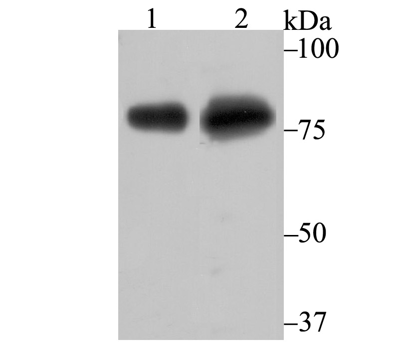
Fig1: Western blot analysis of Tle6 on different lysates using anti-Tle6 antibody at 1/500 dilution. Positive control: Lane1: Mouse ovary tissue Lane2: HepG2Application
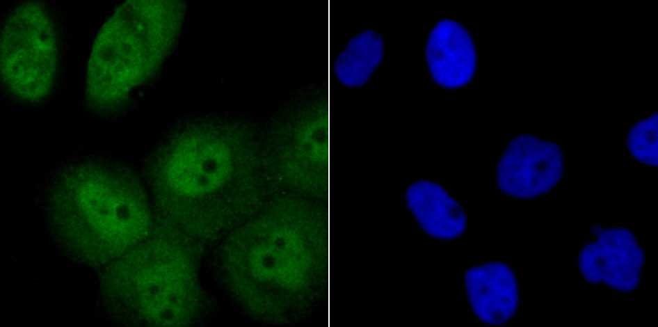
Fig2: ICC staining Tle6 in A431 cells (green). The nuclear counter stain is DAPI (blue). Cells were fixed in paraformaldehyde, permeabilised with 0.25% Triton X100/PBS.Application
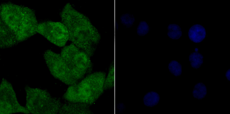
Fig3: ICC staining Tle6 in A549 cells (green). The nuclear counter stain is DAPI (blue). Cells were fixed in paraformaldehyde, permeabilised with 0.25% Triton X100/PBS.Application
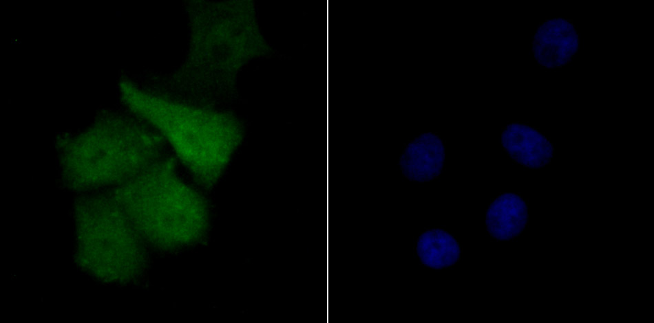
Fig4: ICC staining Tle6 in SK-Br-3 cells (green). The nuclear counter stain is DAPI (blue). Cells were fixed in paraformaldehyde, permeabilised with 0.25% Triton X100/PBS.Application
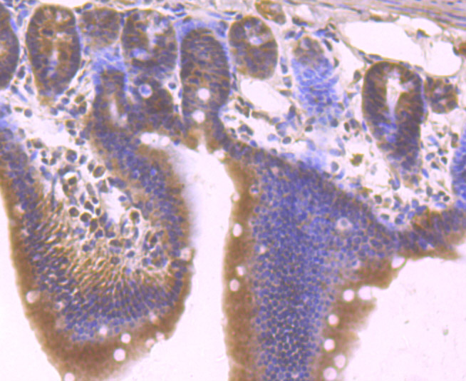
Fig5: Immunohistochemical analysis of paraffin-embedded mouse small intestine tissue using anti-Tle6 antibody. Counter stained with hematoxylin.Application
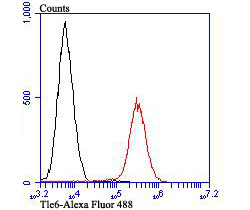
Fig6: Flow cytometric analysis of Daudi cells with Tle6 antibody at 1/100 dilution (red) compared with an unlabelled control (cells without incubation with primary antibody; black). Alexa Fluor 488-conjugated goat anti-rabbit IgG was used as the secondary antibody.| Product Name | Tle6 |
|---|---|
| Antibody Type | Primary Antibodies |
| Product description | The Notch signaling pathway controls various cellular interactions that are important for the specification of a variety of fates in both vertebrates and invertebrates. Key players in the Notch pathway are the TLE genes (for transducin-like enhancer of split, also designated ESG for enhancer of split groucho), which are human homologs of the Drosophila groucho gene. TLE6 (transducin-like enhancer of split 6), also known as GRG6, is a 449 amino acid cytoplasmic protein belonging to the WD repeat groucho/TLE family. As a member of the subcortical maternal complex (SCMC), TLE6 is essential for zygotes to progress beyond the first embryonic cell divisions. TLE6 contains seven WD repeats, a motif known to mediate protein-protein interactions. The WD40 repeat family of proteins is suggested to be involved in signal transduction, RNA processing, gene regulation, vesicular trafficking, cytoskeletal assembly and may play a role in the control of cytotypic differentiation. |
| Immunogen | Synthetic peptide within mouse Tle6 aa 150-200. |
| Clonality | Polyclonal |
|---|---|
| Isotype | IgG |
| Host Species | Rabbit |
| Tested Applications | WBICCIHCFC |
| WB:1:500-1:1,000 ICC:1:50-1:200 IHC:1:50-1:200 FC:1:50-1:100 |
|
| Species Reactivity | HumanMouse |
| Concentration | 1mg/ml |
| Molecular Weight(MW) | 63 kDa |
|---|---|
| Cellular Localization | Nucleus. Cytoplasm. |
Application
Fig1: Western blot analysis of Tle6 on different lysates using anti-Tle6 antibody at 1/500 dilution. Positive control: Lane1: Mouse ovary tissue Lane2: HepG2
Application
Fig2: ICC staining Tle6 in A431 cells (green). The nuclear counter stain is DAPI (blue). Cells were fixed in paraformaldehyde, permeabilised with 0.25% Triton X100/PBS.
Application
Fig3: ICC staining Tle6 in A549 cells (green). The nuclear counter stain is DAPI (blue). Cells were fixed in paraformaldehyde, permeabilised with 0.25% Triton X100/PBS.
Application
Fig4: ICC staining Tle6 in SK-Br-3 cells (green). The nuclear counter stain is DAPI (blue). Cells were fixed in paraformaldehyde, permeabilised with 0.25% Triton X100/PBS.
Application
Fig5: Immunohistochemical analysis of paraffin-embedded mouse small intestine tissue using anti-Tle6 antibody. Counter stained with hematoxylin.
Application
Fig6: Flow cytometric analysis of Daudi cells with Tle6 antibody at 1/100 dilution (red) compared with an unlabelled control (cells without incubation with primary antibody; black). Alexa Fluor 488-conjugated goat anti-rabbit IgG was used as the secondary antibody.| Positive Control | Mouse ovary tissue lysate, HepG2, A431, A549, SK-Br-3, mouse small intestine tissue, Daudi. |
|---|---|
| Application Notes | WB:1:500-1:1,000 ICC:1:50-1:200 IHC:1:50-1:200 FC:1:50-1:100 |
| Form | Liquid |
|---|---|
| Storage Instructions | Store at +4℃ after thawing. Aliquot store at -20℃. Avoid repeated freeze / thaw cycles. |
| Storage Buffer | 1*TBS (pH7.4), 0.5%BSA, 50%Glycerol. Preservative: 0.05% Sodium Azide. |


