Application
Fig4: Flow cytometric analysis of HepG2 cells with SDF1 antibody at 1/100 dilution (red) compared with an unlabelled control (cells without incubation with primary antibody; black). Alexa Fluor 488-conjugated goat anti rabbit IgG was used as the secondary antibody.Application
Fig4: Flow cytometric analysis of HepG2 cells with SDF1 antibody at 1/100 dilution (red) compared with an unlabelled control (cells without incubation with primary antibody; black). Alexa Fluor 488-conjugated goat anti rabbit IgG was used as the secondary antibody.Application
Fig4: Flow cytometric analysis of HepG2 cells with SDF1 antibody at 1/100 dilution (red) compared with an unlabelled control (cells without incubation with primary antibody; black). Alexa Fluor 488-conjugated goat anti rabbit IgG was used as the secondary antibody.Application
Fig4: Flow cytometric analysis of HepG2 cells with SDF1 antibody at 1/100 dilution (red) compared with an unlabelled control (cells without incubation with primary antibody; black). Alexa Fluor 488-conjugated goat anti rabbit IgG was used as the secondary antibody.| Product Name | CXCL12 |
|---|---|
| Antibody Type | Primary Antibodies |
| Antigen Alias | 12-O-tetradecanoylphorbol 13-acetate repressed protein 1 antibody AI174028 antibody C-X-C motif chemokine 12 antibody Chemokine (C-X-C motif) ligand 12 (stromal cell-derived factor 1) antibody Chemokine (C-X-C motif) ligand 12 antibody Chemokine CXC motif ligand 12 antibody cxcl12 antibody hIRH antibody hSDF-1 antibody Intercrine reduced in hepatomas antibody IRH antibody OTTHUMP00000019491 antibody PBSF antibody Pre-B cell growth-stimulating factor antibody SCYB12 antibody SDF 1 antibody SDF-1 antibody SDF-1-alpha(3-67) antibody SDF-1a antibody SDF-1b antibody SDF1_HUMAN antibody SDF1A antibody SDF1B antibody Stromal cell-derived factor 1 antibody Stromal cell-derived factor 1 delta antibody Stromal cell-derived factor 1 gamma antibody Stromal cell-derived factor 1a antibody Stromal cell-derived factor-1 alpha antibody Thymic lymphoma cell-stimulating factor antibody Tlsf antibody TLSF-a antibody TLSF-b antibody Tlsfa antibody Tlsfb antibody TPAR1 antibody |
| Product description | The stromal cell-derived factor 1 (SDF1), also known as C-X-C motif chemokine 12 (CXCL12), is a chemokine protein that in humans is encoded by the CXCL12 gene on chromosome 10. It is ubiquitously expressed in many tissues and cell types. Stromal cell-derived factors 1-alpha and 1-beta are small cytokines that belong to the chemokine family, members of which activate leukocytes and are often induced by proinflammatory stimuli such as lipopolysaccharide, TNF, or IL1. The chemokines are characterized by the presence of 4 conserved cysteines that form 2 disulfide bonds. They can be classified into 2 subfamilies. In the CC subfamily, the cysteine residues are adjacent to each other. In the CXC subfamily, they are separated by an intervening amino acid. The SDF1 proteins belong to the latter group. CXCL12 signaling has been observed in several cancers. The CXCL12 gene also contains one of 27 SNPs associated with increased risk of coronary artery disease. |
| Immunogen | Recombinant protein |
| Clonality | Polyclonal |
|---|---|
| Isotype | IgG |
| Host Species | Rabbit |
| Tested Applications | WBIHCFC |
| WB:1:500 IHC:1:50-1:200 FC:1:50-1:100 |
|
| Species Reactivity | Human |
| Concentration | 1mg/ml |
| Alternative Names | 12-O-tetradecanoylphorbol 13-acetate repressed protein 1 antibody AI174028 antibody C-X-C motif chemokine 12 antibody Chemokine (C-X-C motif) ligand 12 (stromal cell-derived factor 1) antibody Chemokine (C-X-C motif) ligand 12 antibody Chemokine CXC motif ligand 12 antibody cxcl12 antibody hIRH antibody hSDF-1 antibody Intercrine reduced in hepatomas antibody IRH antibody OTTHUMP00000019491 antibody PBSF antibody Pre-B cell growth-stimulating factor antibody SCYB12 antibody SDF 1 antibody SDF-1 antibody SDF-1-alpha(3-67) antibody SDF-1a antibody SDF-1b antibody SDF1_HUMAN antibody SDF1A antibody SDF1B antibody Stromal cell-derived factor 1 antibody Stromal cell-derived factor 1 delta antibody Stromal cell-derived factor 1 gamma antibody Stromal cell-derived factor 1a antibody Stromal cell-derived factor-1 alpha antibody Thymic lymphoma cell-stimulating factor antibody Tlsf antibody TLSF-a antibody TLSF-b antibody Tlsfa antibody Tlsfb antibody TPAR1 antibody |
|---|---|
| Molecular Weight(MW) | 10 kDa |
| Cellular Localization | Secreted. |
| SwissProt ID | P48061 |
|---|
Application
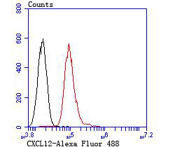
Fig4: Flow cytometric analysis of HepG2 cells with SDF1 antibody at 1/100 dilution (red) compared with an unlabelled control (cells without incubation with primary antibody; black). Alexa Fluor 488-conjugated goat anti rabbit IgG was used as the secondary antibody.
Application
Fig4: Flow cytometric analysis of HepG2 cells with SDF1 antibody at 1/100 dilution (red) compared with an unlabelled control (cells without incubation with primary antibody; black). Alexa Fluor 488-conjugated goat anti rabbit IgG was used as the secondary antibody.
Application
Fig4: Flow cytometric analysis of HepG2 cells with SDF1 antibody at 1/100 dilution (red) compared with an unlabelled control (cells without incubation with primary antibody; black). Alexa Fluor 488-conjugated goat anti rabbit IgG was used as the secondary antibody.
Application
Fig4: Flow cytometric analysis of HepG2 cells with SDF1 antibody at 1/100 dilution (red) compared with an unlabelled control (cells without incubation with primary antibody; black). Alexa Fluor 488-conjugated goat anti rabbit IgG was used as the secondary antibody.| Positive Control | Hela, rat brain tissue, mouse small intestine tissue, HepG2. |
|---|---|
| Application Notes | WB:1:500 IHC:1:50-1:200 FC:1:50-1:100 |
| Form | Liquid |
|---|---|
| Storage Instructions | Store at +4℃ after thawing. Aliquot store at -20℃ or -80℃. Avoid repeated freeze / thaw cycles. |
| Storage Buffer | 1*TBS (pH7.4), 0.5%BSA, 50%Glycerol. Preservative: 0.05% Sodium Azide. |


