Application
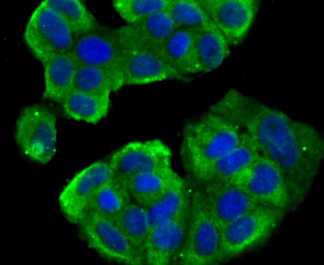
Fig1: ICC staining TWEAKR in MCF-7 cells (green). The nuclear counter stain is DAPI (blue). Cells were fixed in paraformaldehyde, permeabilised with 0.25% Triton X100/PBS.Application
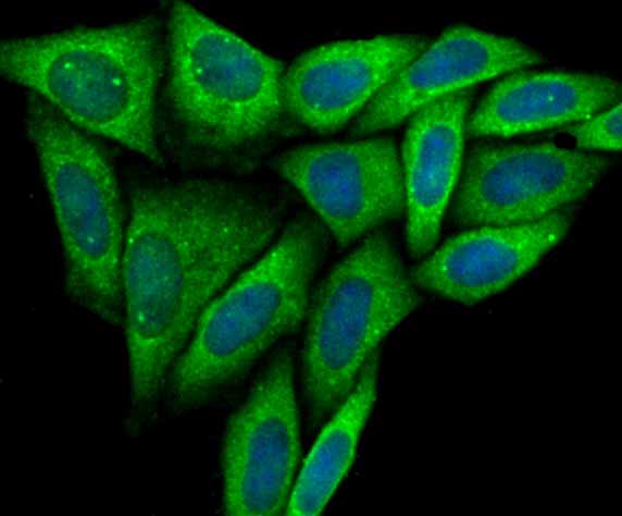
Fig2: ICC staining TWEAKR in SiHa cells (green). The nuclear counter stain is DAPI (blue). Cells were fixed in paraformaldehyde, permeabilised with 0.25% Triton X100/PBS.Application
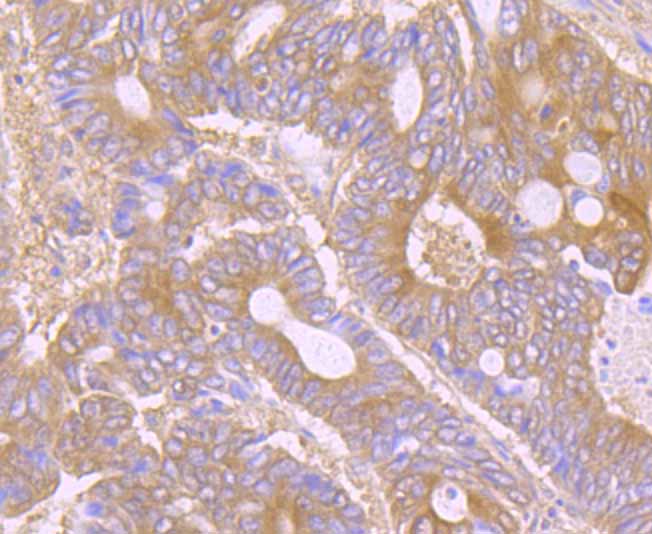
Fig3: Immunohistochemical analysis of paraffin-embedded human colon cancer tissue using anti-TWEAKR antibody. Counter stained with hematoxylin.Application
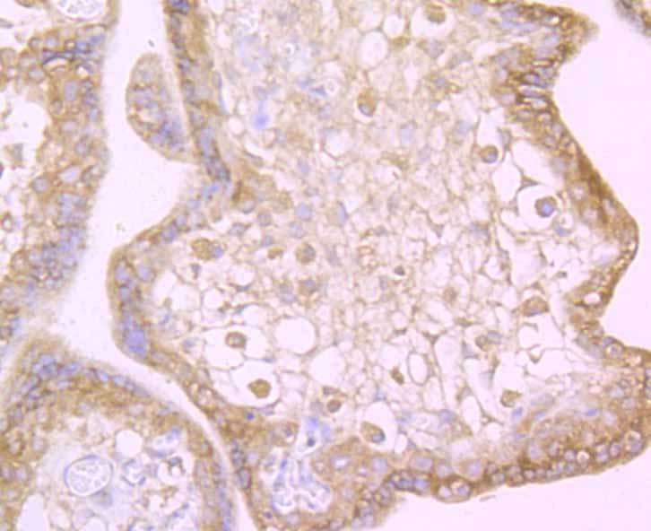
Fig4: Immunohistochemical analysis of paraffin-embedded human placenta tissue using anti-TWEAKR antibody. Counter stained with hematoxylin.Application
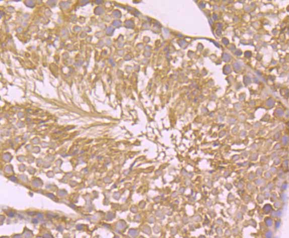
Fig5: Immunohistochemical analysis of paraffin-embedded mouse testis tissue using anti-TWEAKR antibody. Counter stained with hematoxylin.Application
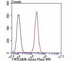
Fig6: Flow cytometric analysis of 293T cells with TWEAKR antibody at 1/100 dilution (red) compared with an unlabelled control (cells without incubation with primary antibody; black). Alexa Fluor 488-conjugated goat anti-rabbit IgG was used as the secondary antibody.| Product Name | TWEAKR |
|---|---|
| Antibody Type | Primary Antibodies |
| Antigen Alias | CD 266 antibody CD266 antibody CD266 antigen antibody FGF inducible 14 antibody FGF-inducible 14 antibody Fibroblast growth factor inducible immediate early response protein 14 antibody Fibroblast growth factor-inducible immediate-early response protein 14 antibody FN 14 antibody FN14 antibody TNFRSF 12A antibody TNFRSF12A antibody TNR12_HUMAN antibody Tumor necrosis factor receptor superfamily member 12A antibody TWEAK R antibody Tweak receptor antibody Tweak-receptor antibody TweakR antibody |
| Product description | Fn14, the TWEAK receptor, is a recently identified member of the TNF receptor superfamily and is expressed on smooth muscle cells and endothelial cells. It is a weak inducer of apoptosis and promotes angiogenesis. Fn14 is a type 1 membrane protein. It associates with TRAF1 and TRAF2, and may modulate cellular adhesion to matrix proteins. Fn14 is highly expressed in heart, placenta and kidney, and moderately expressed in lung, skeletal muscle and pancreas. It is the smallest member of the TNF receptor (TNFR) superfamily described to date, and signals via recruitment of several different TNFR-associated factors. |
| Immunogen | Synthetic peptide within human TWEAKR aa 80-120. |
| Clonality | Polyclonal |
|---|---|
| Isotype | IgG |
| Host Species | Rabbit |
| Tested Applications | ICCIHCFC |
| ICC:1:50-1:200 IHC:1:50-1:200 FC:1:50-1:100 |
|
| Species Reactivity | HumanMouse |
| Concentration | 1mg/ml |
| Alternative Names | CD 266 antibody CD266 antibody CD266 antigen antibody FGF inducible 14 antibody FGF-inducible 14 antibody Fibroblast growth factor inducible immediate early response protein 14 antibody Fibroblast growth factor-inducible immediate-early response protein 14 antibody FN 14 antibody FN14 antibody TNFRSF 12A antibody TNFRSF12A antibody TNR12_HUMAN antibody Tumor necrosis factor receptor superfamily member 12A antibody TWEAK R antibody Tweak receptor antibody Tweak-receptor antibody TweakR antibody |
|---|---|
| Molecular Weight(MW) | 14 kDa |
| Cellular Localization | Membrane. |
Application
Fig1: ICC staining TWEAKR in MCF-7 cells (green). The nuclear counter stain is DAPI (blue). Cells were fixed in paraformaldehyde, permeabilised with 0.25% Triton X100/PBS.
Application
Fig2: ICC staining TWEAKR in SiHa cells (green). The nuclear counter stain is DAPI (blue). Cells were fixed in paraformaldehyde, permeabilised with 0.25% Triton X100/PBS.
Application
Fig3: Immunohistochemical analysis of paraffin-embedded human colon cancer tissue using anti-TWEAKR antibody. Counter stained with hematoxylin.
Application
Fig4: Immunohistochemical analysis of paraffin-embedded human placenta tissue using anti-TWEAKR antibody. Counter stained with hematoxylin.
Application
Fig5: Immunohistochemical analysis of paraffin-embedded mouse testis tissue using anti-TWEAKR antibody. Counter stained with hematoxylin.
Application
Fig6: Flow cytometric analysis of 293T cells with TWEAKR antibody at 1/100 dilution (red) compared with an unlabelled control (cells without incubation with primary antibody; black). Alexa Fluor 488-conjugated goat anti-rabbit IgG was used as the secondary antibody.| Positive Control | MCF-7, SiHa, human colon cancer tissue, human placenta tissue, mouse testis tissue, 293T. |
|---|---|
| Application Notes | ICC:1:50-1:200 IHC:1:50-1:200 FC:1:50-1:100 |
| Form | Liquid |
|---|---|
| Storage Instructions | Store at +4℃ after thawing. Aliquot store at -20℃. Avoid repeated freeze / thaw cycles. |
| Storage Buffer | 1*TBS (pH7.4), 0.5%BSA, 50%Glycerol. Preservative: 0.05% Sodium Azide. |


