Application
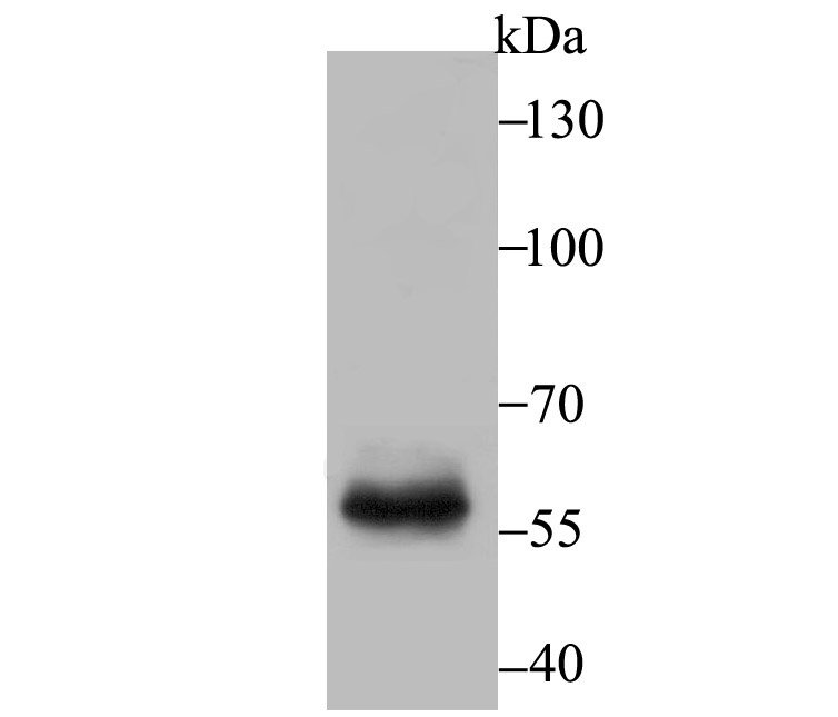
Fig1: Western blot analysis of T-bet on mouse marrow tissue lysate using anti-T-bet antibody at 1/500 dilution.Application
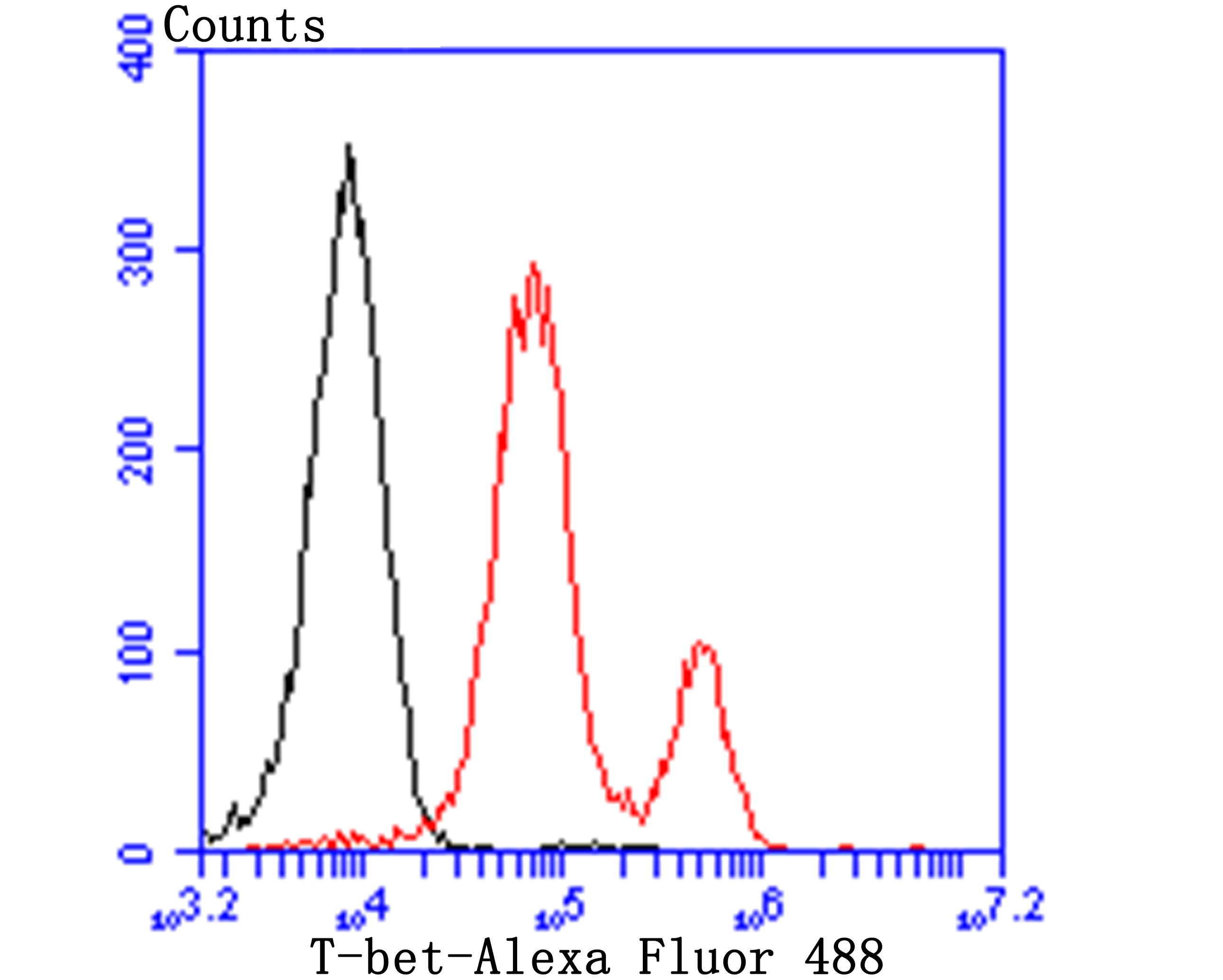
Fig2: Flow cytometric analysis of Jurkat cells with T-bet antibody at 1/100 dilution (red) compared with an unlabelled control (cells without incubation with primary antibody; black). Alexa Fluor 488-conjugated goat anti-rabbit IgG was used as the secondary antibody.| Product Name | T-bet |
|---|---|
| Antibody Type | Primary Antibodies |
| Antigen Alias | T bet antibody T box 21 antibody T box expressed in T cells antibody T box protein 21 antibody T box transcription factor TBX21 antibody T cell specific T box transcription factor antibody T cell specific T box transcription factor T bet antibody T PET antibody T-box protein 21 antibody T-box transcription factor TBX21 antibody T-cell-specific T-box transcription factor T-bet antibody TBET antibody TBLYM antibody TBX 21 antibody Tbx21 antibody TBX21_HUMAN antibody TPET antibody Transcription factor TBLYM antibody |
| Product description | T helper (Th) lymphocytes differentiate into two unique subsets, Th1 and Th2, which differ both in function and in the cytokines they secrete. Th1 and Th2 cytokines promote the growth and differentiation of their subset, and inhibit the growth and differentiation of the opposing subset. T-bet (T box expressed in T cells) is a Th1-specific T box transcription factor that controls the expres-sion of the Th1 cytokine, IFN-γ. T-bet also converts effector Th2 cells into the opposing Th1 subset. T-bet is selectively expressed in Th1 cells. The level of T-bet expression is increased by signals mediated by the T cell receptor (TCR). IL-12 also induces an increase in the level of T-bet. T-bet was originally isolated from nuclear extracts of resting and PMA/ionomycin-activated AE7 cells. T-bet is expressed in low levels in AE7 cells, and in increased levels in stimulated AE7. |
| Immunogen | Synthetic peptide within human T-bet aa 480-530. |
| Clonality | Polyclonal |
|---|---|
| Isotype | IgG |
| Host Species | Rabbit |
| Tested Applications | WBFC |
| WB:1:500 FC:1:50-1:100 |
|
| Species Reactivity | HumanMouse |
| Concentration | 1mg/ml |
| Alternative Names | T bet antibody T box 21 antibody T box expressed in T cells antibody T box protein 21 antibody T box transcription factor TBX21 antibody T cell specific T box transcription factor antibody T cell specific T box transcription factor T bet antibody T PET antibody T-box protein 21 antibody T-box transcription factor TBX21 antibody T-cell-specific T-box transcription factor T-bet antibody TBET antibody TBLYM antibody TBX 21 antibody Tbx21 antibody TBX21_HUMAN antibody TPET antibody Transcription factor TBLYM antibody |
|---|---|
| Molecular Weight(MW) | 58 kDa |
| Cellular Localization | Nucleus. |
| SwissProt ID | Q9JKD8 |
|---|
Application
Fig1: Western blot analysis of T-bet on mouse marrow tissue lysate using anti-T-bet antibody at 1/500 dilution.
Application
Fig2: Flow cytometric analysis of Jurkat cells with T-bet antibody at 1/100 dilution (red) compared with an unlabelled control (cells without incubation with primary antibody; black). Alexa Fluor 488-conjugated goat anti-rabbit IgG was used as the secondary antibody.| Positive Control | Mouse marrow tissue lysate, Jurkat. |
|---|---|
| Application Notes | WB:1:500 FC:1:50-1:100 |
| Form | Liquid |
|---|---|
| Storage Instructions | Store at +4℃ after thawing. Aliquot store at -20℃. Avoid repeated freeze / thaw cycles. |
| Storage Buffer | 1*TBS (pH7.4), 0.5%BSA, 50%Glycerol. Preservative: 0.05% Sodium Azide. |


