Application
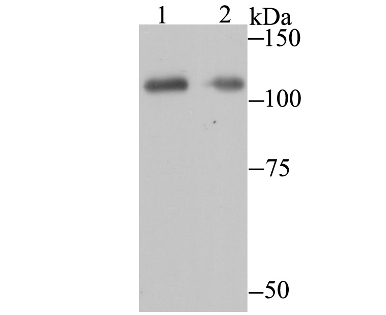
Fig1: Western blot analysis of PARP1 on Daudi and 293 cell lysates using anti- PARP1 antibody at 1/500 dilution.Application
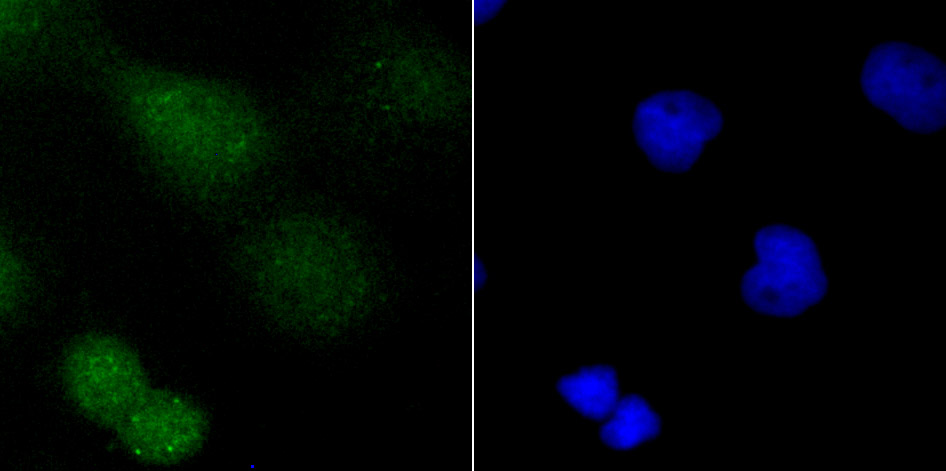
Fig2: ICC staining PARP1 in A431 cells (green). The nuclear counter stain is DAPI (blue). Cells were fixed in paraformaldehyde, permeabilised with 0.25% Triton X100/PBS.Application
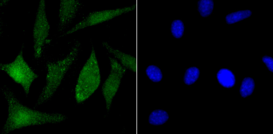
Fig3: ICC staining PARP1 in SH-SY5Y cells (green). The nuclear counter stain is DAPI (blue). Cells were fixed in paraformaldehyde, permeabilised with 0.25% Triton X100/PBS.Application
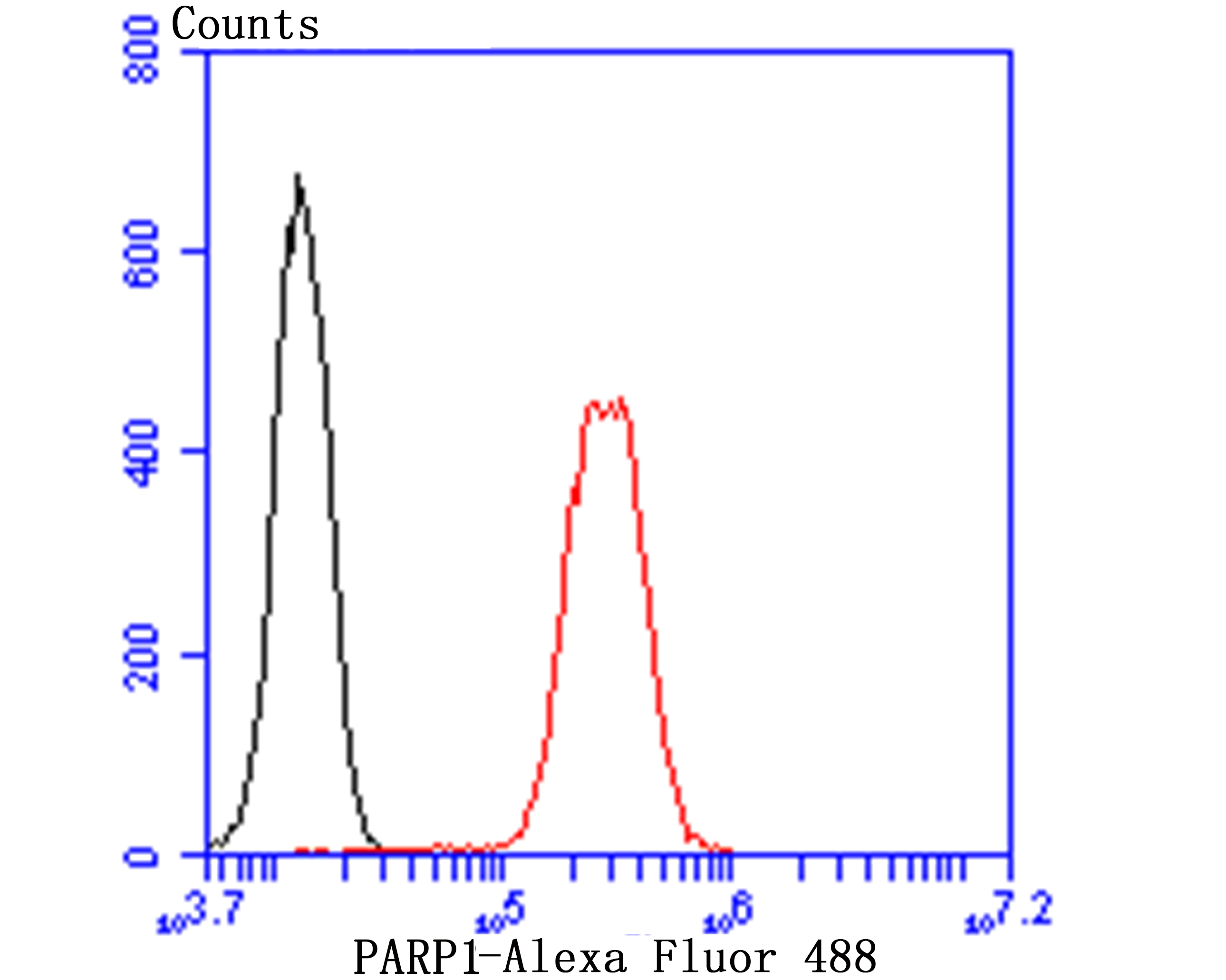
Fig4: Flow cytometric analysis of K562 cells with PARP1 antibody at 1/50 dilution (red) compared with an unlabelled control (cells without incubation with primary antibody; black). Alexa Fluor 488-conjugated goat anti rabbit IgG was used as the secondary antibody.| Product Name | PARP1 |
|---|---|
| Antibody Type | Primary Antibodies |
| Antigen Alias | ADP ribosyltransferase (NAD+; poly (ADP ribose) polymerase) antibody ADP ribosyltransferase antibody ADP ribosyltransferase diphtheria toxin like 1 antibody ADP ribosyltransferase NAD(+) antibody ADPRT 1 antibody ADPRT antibody ADPRT1 antibody ARTD1 antibody msPARP antibody NAD(+) ADP ribosyltransferase 1 antibody NAD(+) ADP-ribosyltransferase 1 antibody pADPRT 1 antibody pADPRT1 antibody PARP 1 antibody PARP antibody PARP-1 antibody PARP1 antibody PARP1_HUMAN antibody Poly (ADP ribose) polymerase 1 antibody poly (ADP ribose) polymerase family, member 1 antibody Poly [ADP-ribose] polymerase 1 antibody Poly(ADP ribose) polymerase antibody poly(ADP ribose) synthetase antibody poly(ADP ribosyl)transferase antibody Poly[ADP ribose] synthetase 1 antibody Poly[ADP-ribose] synthase 1 antibody PPOL antibody |
| Product description | Involved in the base excision repair (BER) pathway, by catalyzing the poly(ADP-ribosyl)ation of a limited number of acceptor proteins involved in chromatin architecture and in DNA metabolism. This modification follows DNA damages and appears as an obligatory step in a detection/signaling pathway leading to the reparation of DNA strand breaks. Mediates the poly(ADP-ribosyl)ation of APLF and CHFR. Positively regulates the transcription of MTUS1 and negatively regulates the transcription of MTUS2/TIP150. With EEF1A1 and TXK, forms a complex that acts as a T-helper 1 (Th1) cell-specific transcription factor and binds the promoter of IFN-gamma to directly regulate its transcription, and is thus involved importantly in Th1 cytokine production. Required for PARP9 and DTX3L recruitment to DNA damage sites. PARP1-dependent PARP9-DTX3L-mediated ubiquitination promotes the rapid and specific recruitment of 53BP1/TP53BP1, UIMC1/RAP80, and BRCA1 to DNA damage sites. |
| Immunogen | Synthetic peptide within PARP1 aa 200-250. |
| Clonality | Polyclonal |
|---|---|
| Isotype | IgG |
| Host Species | Rabbit |
| Tested Applications | WBICCFC |
| WB:1:500 ICC:1:50-1:100 FC:1:50-1:100 |
|
| Species Reactivity | HumanMouse |
| Concentration | 1mg/ml |
| Alternative Names | ADP ribosyltransferase (NAD+ poly (ADP ribose) polymerase) antibody ADP ribosyltransferase antibody ADP ribosyltransferase diphtheria toxin like 1 antibody ADP ribosyltransferase NAD(+) antibody ADPRT 1 antibody ADPRT antibody ADPRT1 antibody ARTD1 antibody msPARP antibody NAD(+) ADP ribosyltransferase 1 antibody NAD(+) ADP-ribosyltransferase 1 antibody pADPRT 1 antibody pADPRT1 antibody PARP 1 antibody PARP antibody PARP-1 antibody PARP1 antibody PARP1_HUMAN antibody Poly (ADP ribose) polymerase 1 antibody poly (ADP ribose) polymerase family member 1 antibody Poly [ADP-ribose] polymerase 1 antibody Poly(ADP ribose) polymerase antibody poly(ADP ribose) synthetase antibody poly(ADP ribosyl)transferase antibody Poly[ADP ribose] synthetase 1 antibody Poly[ADP-ribose] synthase 1 antibody PPOL antibody |
|---|---|
| Molecular Weight(MW) | 113 kDa |
| Cellular Localization | Nucleus. |
| SwissProt ID | P09874 |
|---|
Application
Fig1: Western blot analysis of PARP1 on Daudi and 293 cell lysates using anti- PARP1 antibody at 1/500 dilution.
Application
Fig2: ICC staining PARP1 in A431 cells (green). The nuclear counter stain is DAPI (blue). Cells were fixed in paraformaldehyde, permeabilised with 0.25% Triton X100/PBS.
Application
Fig3: ICC staining PARP1 in SH-SY5Y cells (green). The nuclear counter stain is DAPI (blue). Cells were fixed in paraformaldehyde, permeabilised with 0.25% Triton X100/PBS.
Application
Fig4: Flow cytometric analysis of K562 cells with PARP1 antibody at 1/50 dilution (red) compared with an unlabelled control (cells without incubation with primary antibody; black). Alexa Fluor 488-conjugated goat anti rabbit IgG was used as the secondary antibody.| Positive Control | Daudi, 293, A431, PC-3M, K562. |
|---|---|
| Application Notes | WB:1:500 ICC:1:50-1:100 FC:1:50-1:100 |
| Form | Liquid |
|---|---|
| Storage Instructions | Store at +4℃ after thawing. Aliquot store at -20℃. Avoid repeated freeze / thaw cycles. |
| Storage Buffer | 1*TBS (pH7.4), 0.5%BSA, 50%Glycerol. Preservative: 0.05% Sodium Azide. |


