Application
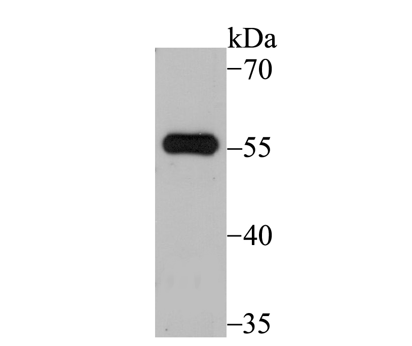
Fig1: Western blot analysis of SUFU on 293 cell lysate using anti-SUFU antibody at 1/500 dilution.Application
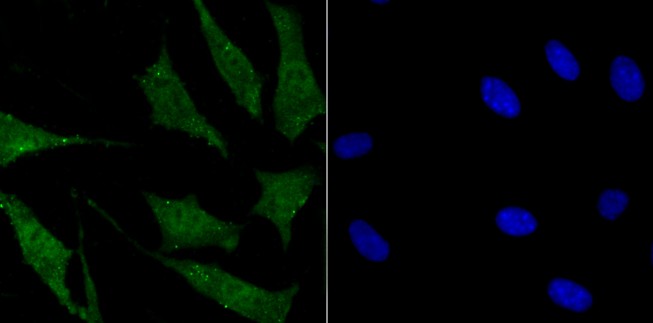
Fig2: ICC staining SUFU in SH-SY-5Y cells (green). The nuclear counter stain is DAPI (blue). Cells were fixed in paraformaldehyde, permeabilised with 0.25% Triton X100/PBS.Application
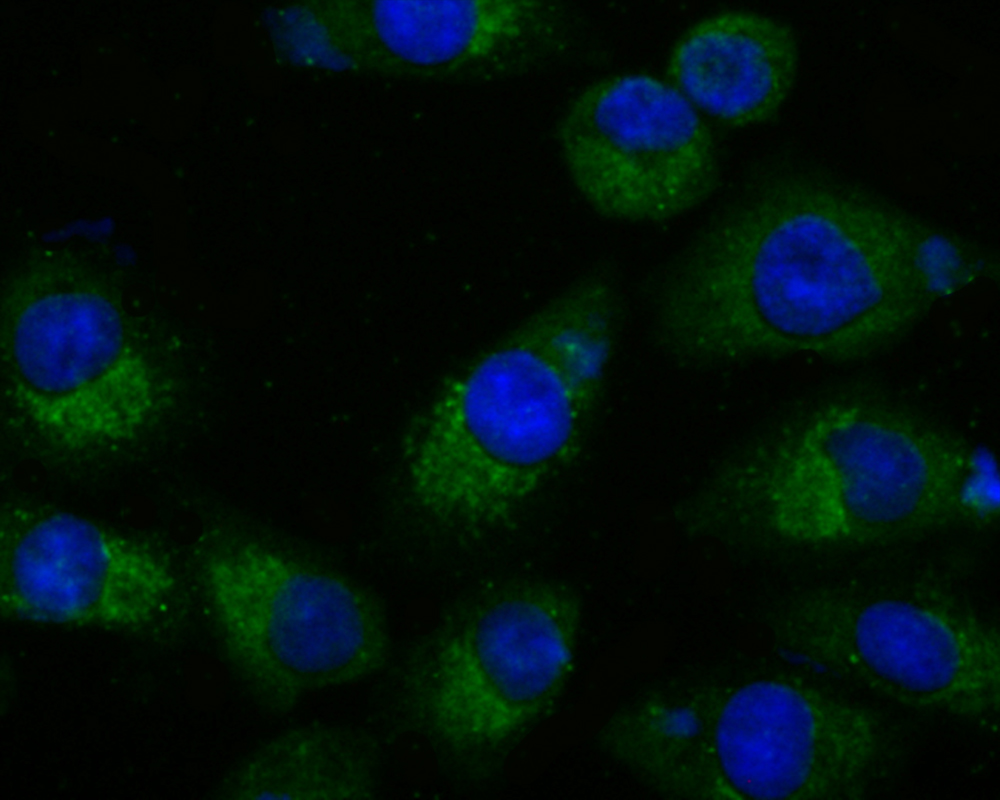
Fig3: ICC staining SUFU in A549 cells (green). The nuclear counter stain is DAPI (blue). Cells were fixed in paraformaldehyde, permeabilised with 0.25% Triton X100/PBS.Application
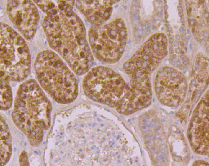
Fig4: Immunohistochemical analysis of paraffin-embedded human kidney tissue using anti-SUFU antibody. Counter stained with hematoxylin.Application
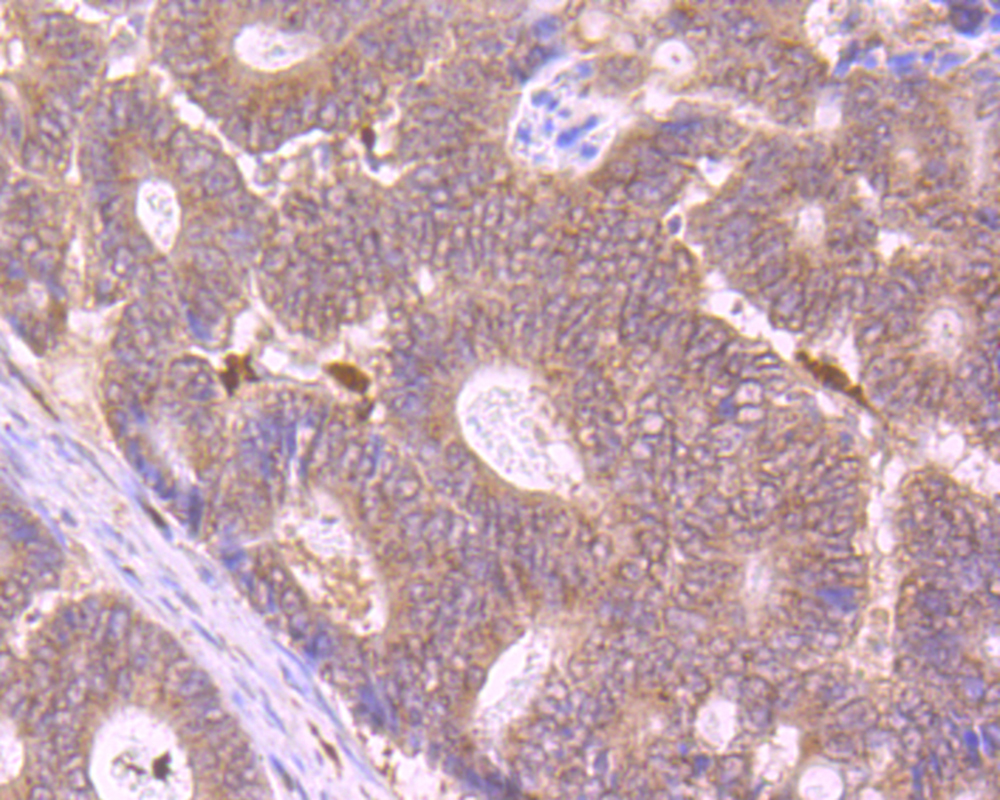
Fig5: Immunohistochemical analysis of paraffin-embedded human colon cancer tissue using anti-SUFU antibody. Counter stained with hematoxylin.| Product Name | SUFU |
|---|---|
| Antibody Type | Primary Antibodies |
| Antigen Alias | OTTHUMP00000020374 antibody OTTHUMP00000020377 antibody OTTHUMP00000020379 antibody PRO1280 antibody SU FU antibody SU(F)U antibody Su(fu) antibody SUFU antibody SUFU negative regulator of hedgehog signaling antibody SUFU_HUMAN antibody SUFUH antibody SUFUXL antibody Suppressor of fused homolog (Drosophila) antibody Suppressor of fused homolog antibody |
| Product description | Su(fu) (for Suppressor-of-Fused) is a key negative regulator in the vertebrate Hedgehog signaling pathway. Su(fu) interacts with genes encoding proteins in this signal transduction pathway. In Drosophila, Intracellular transduction of the Hedgehog pathway involves the release of a large complex containing Su(fu). Su(fu) inhibits the activity of the transcription factor Gli1 and interacts with Gli2, Gli3 and the serine/threonine kinase Fused. Su(fu) is widely expressed in adult and embryonic tissues with higher expression in tissues patterned by hegdehog signaling. The Su(fu) gene locus maps to a region that is deleted in glioblastomas, prostate cancer, malignant melanoma and endometrial cancer. |
| Immunogen | Recombinant protein with human SUFU aa 1-250. |
| Clonality | Polyclonal |
|---|---|
| Isotype | IgG |
| Host Species | Rabbit |
| Tested Applications | WBICCIHC |
| WB:1:500-1:1,000 ICC:1:50-1:200 IHC:1:50-1:200 |
|
| Species Reactivity | Human |
| Concentration | 1mg/ml |
| Alternative Names | OTTHUMP00000020374 antibody OTTHUMP00000020377 antibody OTTHUMP00000020379 antibody PRO1280 antibody SU FU antibody SU(F)U antibody Su(fu) antibody SUFU antibody SUFU negative regulator of hedgehog signaling antibody SUFU_HUMAN antibody SUFUH antibody SUFUXL antibody Suppressor of fused homolog (Drosophila) antibody Suppressor of fused homolog antibody |
|---|---|
| Molecular Weight(MW) | 54 kDa |
| Cellular Localization | Cytoplasm. Nucleus. |
| SwissProt ID | Q9UMX1 |
|---|
Application
Fig1: Western blot analysis of SUFU on 293 cell lysate using anti-SUFU antibody at 1/500 dilution.
Application
Fig2: ICC staining SUFU in SH-SY-5Y cells (green). The nuclear counter stain is DAPI (blue). Cells were fixed in paraformaldehyde, permeabilised with 0.25% Triton X100/PBS.
Application
Fig3: ICC staining SUFU in A549 cells (green). The nuclear counter stain is DAPI (blue). Cells were fixed in paraformaldehyde, permeabilised with 0.25% Triton X100/PBS.
Application
Fig4: Immunohistochemical analysis of paraffin-embedded human kidney tissue using anti-SUFU antibody. Counter stained with hematoxylin.
Application
Fig5: Immunohistochemical analysis of paraffin-embedded human colon cancer tissue using anti-SUFU antibody. Counter stained with hematoxylin.| Positive Control | 293, SH-SY-5Y, A549, human kidney tissue, human colon cancer tissue. |
|---|---|
| Application Notes | WB:1:500-1:1,000 ICC:1:50-1:200 IHC:1:50-1:200 |
| Form | Liquid |
|---|---|
| Storage Instructions | Store at +4℃ after thawing. Aliquot store at -20℃. Avoid repeated freeze / thaw cycles. |
| Storage Buffer | 1*TBS (pH7.4), 0.5%BSA, 50%Glycerol. Preservative: 0.05% Sodium Azide. |


