Application
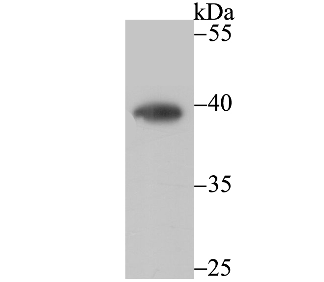
Fig1: Western blot analysis of CTGF on mouse smooth muscle tissue lysate using anti-CTGF antibody at 1/500 dilution.Application
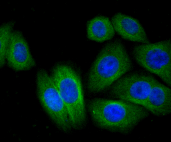
Fig2: ICC staining CTGF in HepG2 cells (green). The nuclear counter stain is DAPI (blue). Cells were fixed in paraformaldehyde, permeabilised with 0.25% Triton X100/PBS.Application
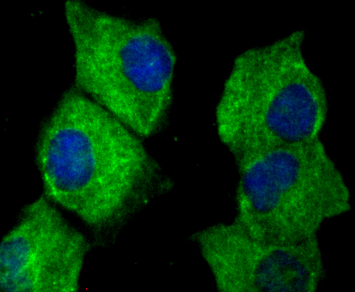
Fig3: ICC staining CTGF in HUVEC cells (green). The nuclear counter stain is DAPI (blue). Cells were fixed in paraformaldehyde, permeabilised with 0.25% Triton X100/PBS.Application
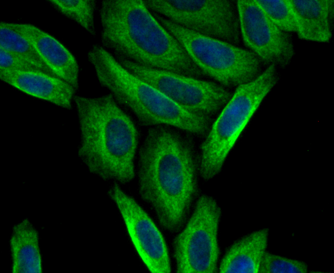
Fig4: ICC staining CTGF in SiHa cells (green). The nuclear counter stain is DAPI (blue). Cells were fixed in paraformaldehyde, permeabilised with 0.25% Triton X100/PBS.Application
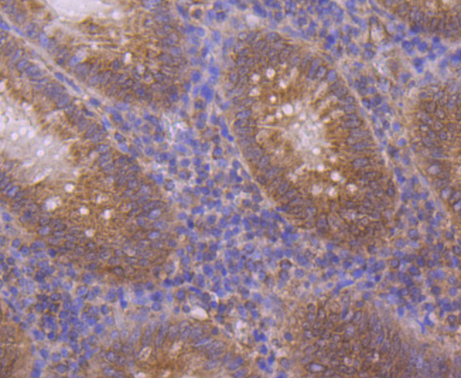
Fig5: Immunohistochemical analysis of paraffin-embedded human appendix tissue using anti-CTGF antibody. Counter stained with hematoxylin.Application
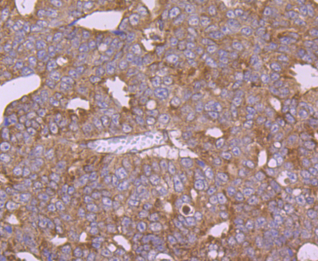
Fig6: Immunohistochemical analysis of paraffin-embedded human thyroid gland cancer tissue using anti-CTGF antibody. Counter stained with hematoxylin.Application
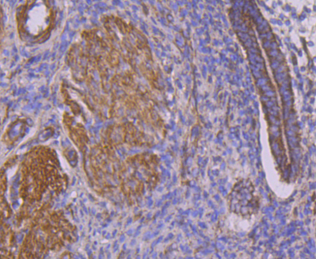
Fig7: Immunohistochemical analysis of paraffin-embedded human uterus tissue using anti-CTGF antibody. Counter stained with hematoxylin.| Product Name | CTGF |
|---|---|
| Antibody Type | Primary Antibodies |
| Antigen Alias | CCN 2 antibody CCN family member 2 antibody CCN2 antibody Connective tissue growth factor antibody Ctgf antibody CTGF_HUMAN antibody Hcs 24 antibody Hcs24 antibody Hypertrophic chondrocyte specific protein 24 antibody Hypertrophic chondrocyte-specific gene product 24 antibody Hypertrophic chondrocyte-specific protein 24 antibody IBP-8 antibody IGF-binding protein 8 antibody IGFBP-8 antibody IGFBP8 antibody Insulin like Growth Factor Binding Protein 8 antibody Insulin-like growth factor-binding protein 8 antibody MGC102839 antibody NOV 2 antibody NOV2 antibody |
| Product description | Connective tissue growth factor (CTGF, also known as hypertrophic chondrocyte-specific gene product 24 or Hcs24), is a member of the CCN family of immediate early proteins, which are involved in cell proliferation, migration and matrix production. CTGF is a cysteine-rich peptide that is secreted by endothelial cells, fibroblasts, smooth muscle cells and myofibroblasts. Its expression is increased in various human and animal fibrotic diseases. Specifically, CTGF has been observed to be strongly upregulated in human proliferative and fibrogenic renal disease. In addition, CTGF is a growth factor for vascular smooth muscle cells (VSMC) and may play a similar role in promoting VSMC growth and migration in vitro. |
| Immunogen | Recombinant protein with human CTGF aa 150-300. |
| Clonality | Polyclonal |
|---|---|
| Isotype | IgG |
| Host Species | Rabbit |
| Tested Applications | WBICCIHC |
| WB:1:500 ICC:1:50-1:200 IHC:1:50-1:200 |
|
| Species Reactivity | HumanMouse |
| Concentration | 1mg/ml |
| Alternative Names | CCN 2 antibody CCN family member 2 antibody CCN2 antibody Connective tissue growth factor antibody Ctgf antibody CTGF_HUMAN antibody Hcs 24 antibody Hcs24 antibody Hypertrophic chondrocyte specific protein 24 antibody Hypertrophic chondrocyte-specific gene product 24 antibody Hypertrophic chondrocyte-specific protein 24 antibody IBP-8 antibody IGF-binding protein 8 antibody IGFBP-8 antibody IGFBP8 antibody Insulin like Growth Factor Binding Protein 8 antibody Insulin-like growth factor-binding protein 8 antibody MGC102839 antibody NOV 2 antibody NOV2 antibody |
|---|---|
| Molecular Weight(MW) | 38 kDa |
| Cellular Localization | Extracellular region or secreted. |
Application
Fig1: Western blot analysis of CTGF on mouse smooth muscle tissue lysate using anti-CTGF antibody at 1/500 dilution.
Application
Fig2: ICC staining CTGF in HepG2 cells (green). The nuclear counter stain is DAPI (blue). Cells were fixed in paraformaldehyde, permeabilised with 0.25% Triton X100/PBS.
Application
Fig3: ICC staining CTGF in HUVEC cells (green). The nuclear counter stain is DAPI (blue). Cells were fixed in paraformaldehyde, permeabilised with 0.25% Triton X100/PBS.
Application
Fig4: ICC staining CTGF in SiHa cells (green). The nuclear counter stain is DAPI (blue). Cells were fixed in paraformaldehyde, permeabilised with 0.25% Triton X100/PBS.
Application
Fig5: Immunohistochemical analysis of paraffin-embedded human appendix tissue using anti-CTGF antibody. Counter stained with hematoxylin.
Application
Fig6: Immunohistochemical analysis of paraffin-embedded human thyroid gland cancer tissue using anti-CTGF antibody. Counter stained with hematoxylin.
Application
Fig7: Immunohistochemical analysis of paraffin-embedded human uterus tissue using anti-CTGF antibody. Counter stained with hematoxylin.| Positive Control | Mouse smooth muscle tissue lysate, HepG2, HUVEC, SiHa, human appendix tissue, human thyroid gland cancer tissue, human uterus tissue. |
|---|---|
| Application Notes | WB:1:500 ICC:1:50-1:200 IHC:1:50-1:200 |
| Form | Liquid |
|---|---|
| Storage Instructions | Store at +4℃ after thawing. Aliquot store at -20℃. Avoid repeated freeze / thaw cycles. |
| Storage Buffer | 1*TBS (pH7.4), 0.5%BSA, 50%Glycerol. Preservative: 0.05% Sodium Azide. |


