Application
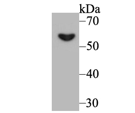
Fig1: Western blot analysis of PKM2 on 293 cell lysate using anti-PKM2 antibody at 1/500 dilution.Application
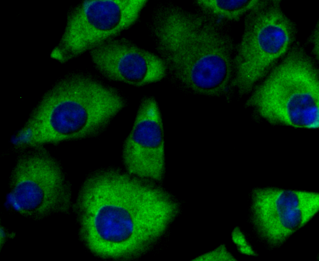
Fig2: ICC staining PKM2 in A549 cells (green). The nuclear counter stain is DAPI (blue). Cells were fixed in paraformaldehyde, permeabilised with 0.25% Triton X100/PBS.Application
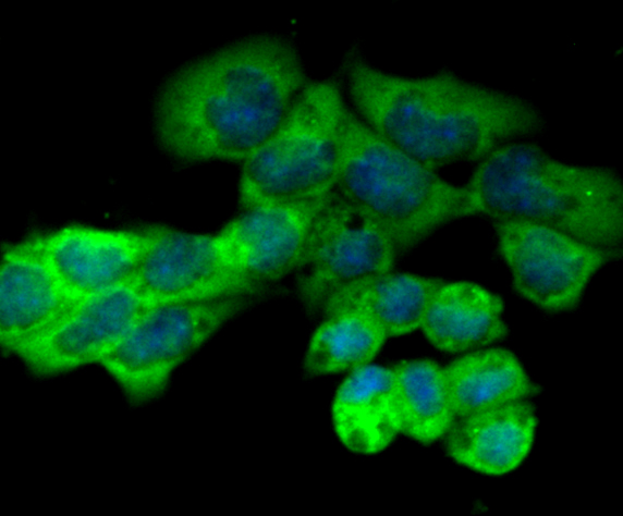
Fig3: ICC staining PKM2 in F9 cells (green). The nuclear counter stain is DAPI (blue). Cells were fixed in paraformaldehyde, permeabilised with 0.25% Triton X100/PBS.Application
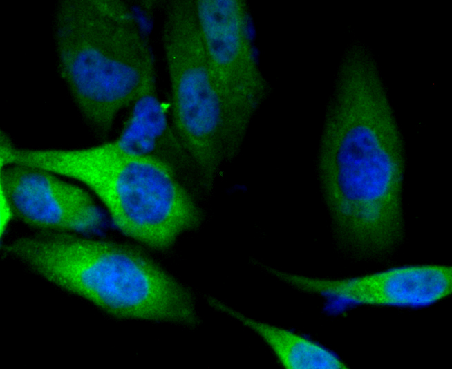
Fig4: ICC staining PKM2 in PC-3M cells (green). The nuclear counter stain is DAPI (blue). Cells were fixed in paraformaldehyde, permeabilised with 0.25% Triton X100/PBS.Application
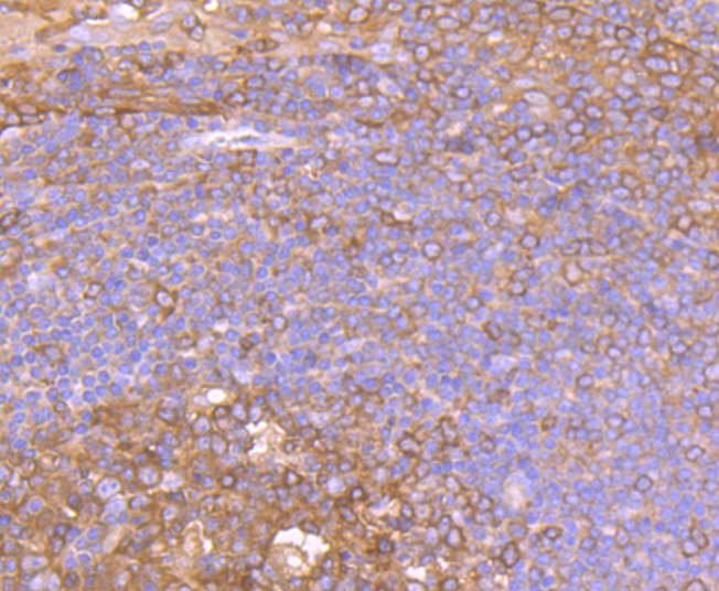
Fig5: Immunohistochemical analysis of paraffin-embedded human tonsil tissue using anti-PKM2 antibody. Counter stained with hematoxylin.Application
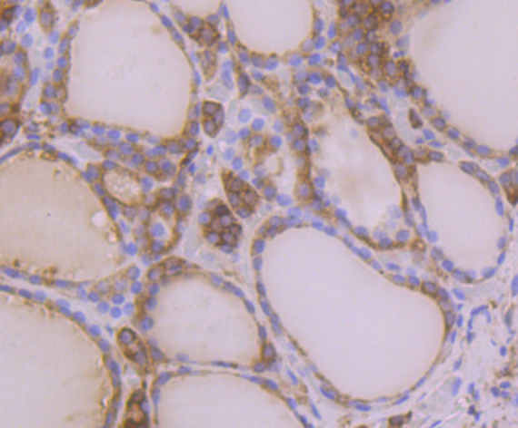
Fig6: Immunohistochemical analysis of paraffin-embedded human thyroid gland cancer tissue using anti-PKM2 antibody. Counter stained with hematoxylin.Application
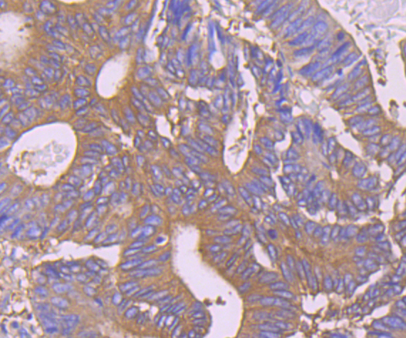
Fig7: Immunohistochemical analysis of paraffin-embedded human colon cancer tissue using anti-PKM2 antibody. Counter stained with hematoxylin.Application
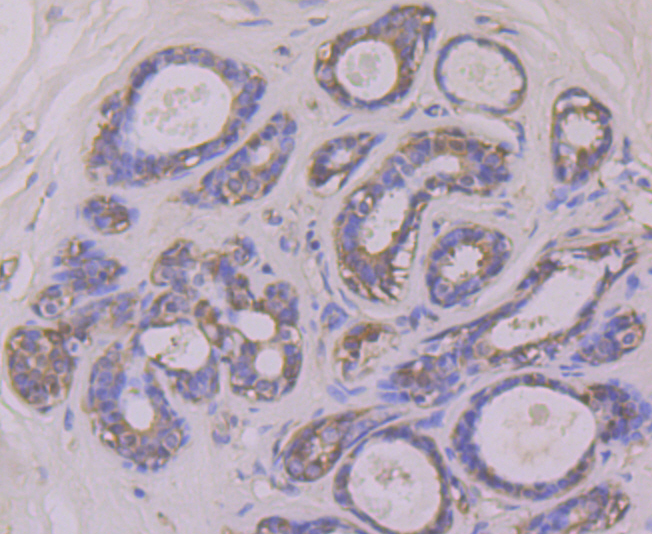
Fig8: Immunohistochemical analysis of paraffin-embedded human breast cancer tissue using anti-PKM2 antibody. Counter stained with hematoxylin.| Product Name | PKM2 |
|---|---|
| Antibody Type | Primary Antibodies |
| Antigen Alias | CTHBP antibody Cytosolic thyroid hormone binding protein antibody Cytosolic thyroid hormone-binding protein antibody KPYM_HUMAN antibody MGC3932 antibody OIP 3 antibody OIP-3 antibody OIP3 antibody OPA interacting protein 3 antibody Opa-interacting protein 3 antibody p58 antibody PK muscle type antibody PK, muscle type antibody PK2 antibody PK3 antibody PKM antibody PKM2 antibody pykm antibody Pyruvate kinase 2/3 antibody Pyruvate kinase 3 antibody Pyruvate kinase isozymes M1/M2 antibody Pyruvate kinase muscle antibody Pyruvate kinase muscle isozyme antibody pyruvate kinase PKM antibody Pyruvate kinase, muscle 2 antibody TCB antibody THBP1 antibody Thyroid hormone binding protein 1 antibody Thyroid hormone binding protein cytosolic antibody Thyroid hormone-binding protein 1 antibody Tumor M2 PK antibody Tumor M2-PK antibody |
| Product description | In mammals, four different isoenzymes exist for pyruvate kinase. Pyruvate kinases are responsible for catalyzing the final step in glycolysis: the conversion of phosphoenolpyruvate to pyruvate with the coinciding generation of ATP. The PKM (pyruvate kinase, muscle) gene encodes the M1- and M2-type isoenzymes through alternative splicing events. Both M1- and M2-type isoforms exists as tetramers and are stimulated by fructose 1,6-bisphosphate. In addition, both isoforms exhibit thyroid hormone binding activity and may be referred to as CTHBP (cytosolic thyroid hormone-binding protein) or THBP1. The M2-type isoform also interacts with Oct-4 via its C-terminal domain, functioning to enhance Oct-4 transcriptional activity. Translocates to the nucleus in response to different apoptotic stimuli. Nuclear translocation is sufficient to induce cell death that is caspase independent, isoform-specific and independent of its enzymatic activity. |
| Immunogen | Synthetic peptide within human PKM2 aa 380-430. |
| Clonality | Polyclonal |
|---|---|
| Isotype | IgG |
| Host Species | Rabbit |
| Tested Applications | WBICCIHC |
| WB:1:500-1:1,000 ICC:1:100-1:500 IHC:1:50-1:200 |
|
| Species Reactivity | Human |
| Concentration | 1mg/ml |
| Alternative Names | CTHBP antibody
Cytosolic thyroid hormone binding protein antibody
Cytosolic thyroid hormone-binding protein antibody
KPYM_HUMAN antibody
MGC3932 antibody
OIP 3 antibody
OIP-3 antibody
OIP3 antibody
OPA interacting protein 3 antibody
Opa-interacting protein 3 antibody
p58 antibody
PK muscle type antibody
PK muscle type antibody PK2 antibody PK3 antibody PKM antibody PKM2 antibody pykm antibody Pyruvate kinase 2/3 antibody Pyruvate kinase 3 antibody Pyruvate kinase isozymes M1/M2 antibody Pyruvate kinase muscle antibody Pyruvate kinase muscle isozyme antibody pyruvate kinase PKM antibody Pyruvate kinase muscle 2 antibody TCB antibody THBP1 antibody Thyroid hormone binding protein 1 antibody Thyroid hormone binding protein cytosolic antibody Thyroid hormone-binding protein 1 antibody Tumor M2 PK antibody Tumor M2-PK antibody |
|---|---|
| Molecular Weight(MW) | 58 kDa |
| Cellular Localization | Cytoplasm. Nucleus. |
| SwissProt ID | P14618 |
|---|
Application
Fig1: Western blot analysis of PKM2 on 293 cell lysate using anti-PKM2 antibody at 1/500 dilution.
Application
Fig2: ICC staining PKM2 in A549 cells (green). The nuclear counter stain is DAPI (blue). Cells were fixed in paraformaldehyde, permeabilised with 0.25% Triton X100/PBS.
Application
Fig3: ICC staining PKM2 in F9 cells (green). The nuclear counter stain is DAPI (blue). Cells were fixed in paraformaldehyde, permeabilised with 0.25% Triton X100/PBS.
Application
Fig4: ICC staining PKM2 in PC-3M cells (green). The nuclear counter stain is DAPI (blue). Cells were fixed in paraformaldehyde, permeabilised with 0.25% Triton X100/PBS.
Application
Fig5: Immunohistochemical analysis of paraffin-embedded human tonsil tissue using anti-PKM2 antibody. Counter stained with hematoxylin.
Application
Fig6: Immunohistochemical analysis of paraffin-embedded human thyroid gland cancer tissue using anti-PKM2 antibody. Counter stained with hematoxylin.
Application
Fig7: Immunohistochemical analysis of paraffin-embedded human colon cancer tissue using anti-PKM2 antibody. Counter stained with hematoxylin.
Application
Fig8: Immunohistochemical analysis of paraffin-embedded human breast cancer tissue using anti-PKM2 antibody. Counter stained with hematoxylin.| Positive Control | 293, A549, F9, PC-3M, human tonsil tissue, human thyroid gland cancer tissue, human colon cancer tissue, human breast cancer tissue. |
|---|---|
| Application Notes | WB:1:500-1:1,000 ICC:1:100-1:500 IHC:1:50-1:200 |
| Form | Liquid |
|---|---|
| Storage Instructions | Store at +4℃ after thawing. Aliquot store at -20℃. Avoid repeated freeze / thaw cycles. |
| Storage Buffer | 1*TBS (pH7.4), 0.5%BSA, 50%Glycerol. Preservative: 0.05% Sodium Azide. |


