Application
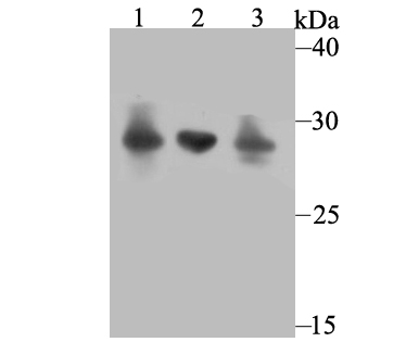
Fig1: Western blot analysis of Carbonic anhydrase 2 on different cell lysates using anti-Carbonic anhydrase 2 antibody at 1/1,000 dilution. Positive control: Lane1: 293 Lane2: THP-1 Lane3: HL-60Application
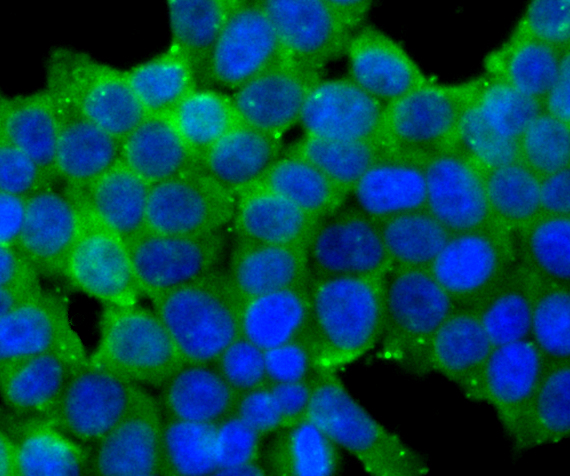
Fig2: ICC staining Carbonic anhydrase 2 in 293T cells (green). The nuclear counter stain is DAPI (blue). Cells were fixed in paraformaldehyde, permeabilised with 0.25% Triton X100/PBS.Application
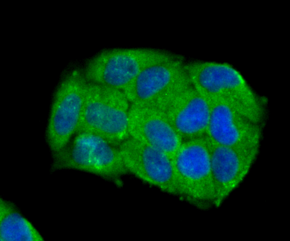
Fig3: ICC staining Carbonic anhydrase 2 in Hela cells (green). The nuclear counter stain is DAPI (blue). Cells were fixed in paraformaldehyde, permeabilised with 0.25% Triton X100/PBS.Application
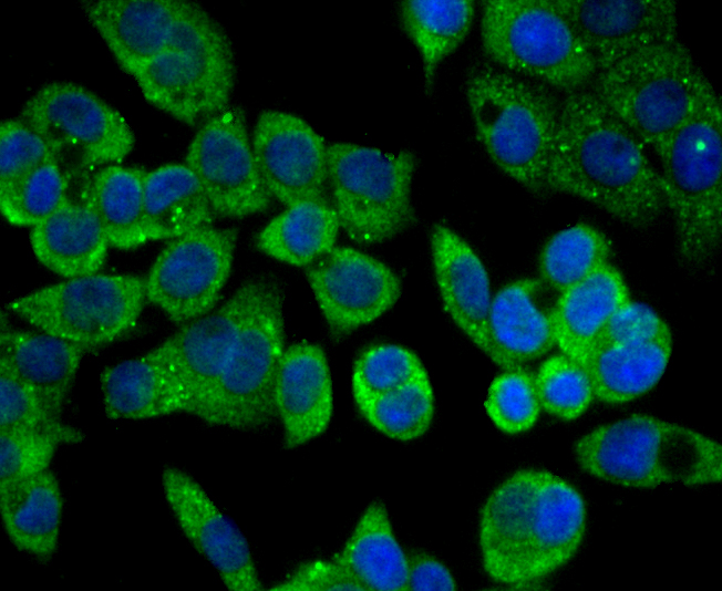
Fig4: ICC staining Carbonic anhydrase 2 in LOVO cells (green). The nuclear counter stain is DAPI (blue). Cells were fixed in paraformaldehyde, permeabilised with 0.25% Triton X100/PBS.Application
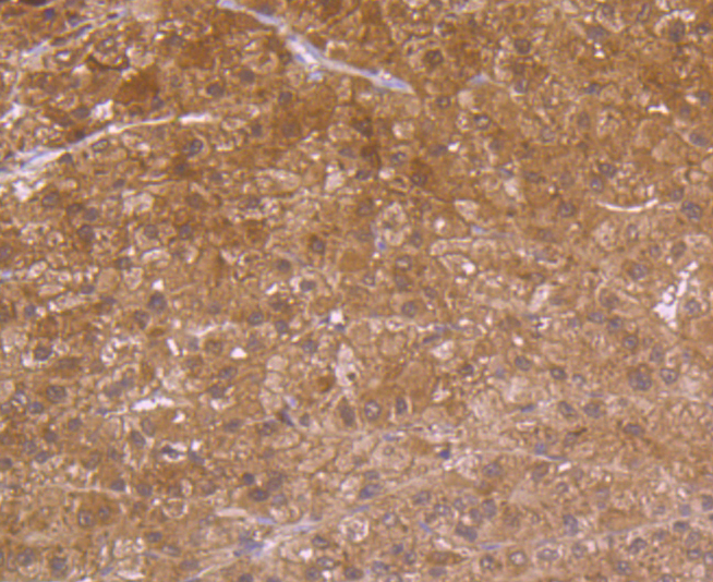
Fig5: Immunohistochemical analysis of paraffin-embedded human liver tissue using anti-Carbonic anhydrase 2 antibody. Counter stained with hematoxylin.Application
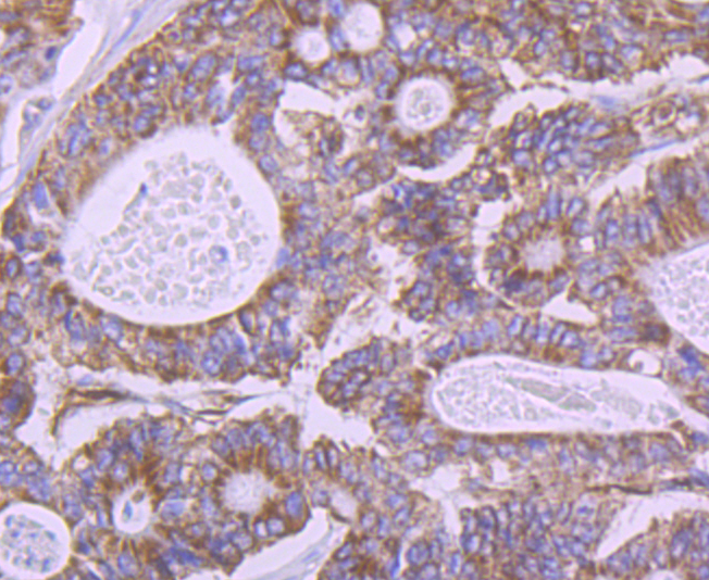
Fig6: Immunohistochemical analysis of paraffin-embedded human colon cancer tissue using anti-Carbonic anhydrase 2 antibody. Counter stained with hematoxylin.Application
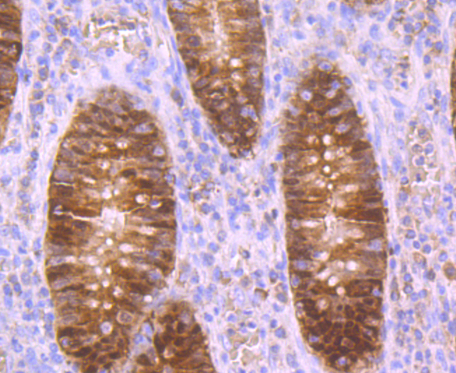
Fig7: Immunohistochemical analysis of paraffin-embedded human appendix tissue using anti-Carbonic anhydrase 2 antibody. Counter stained with hematoxylin.Application
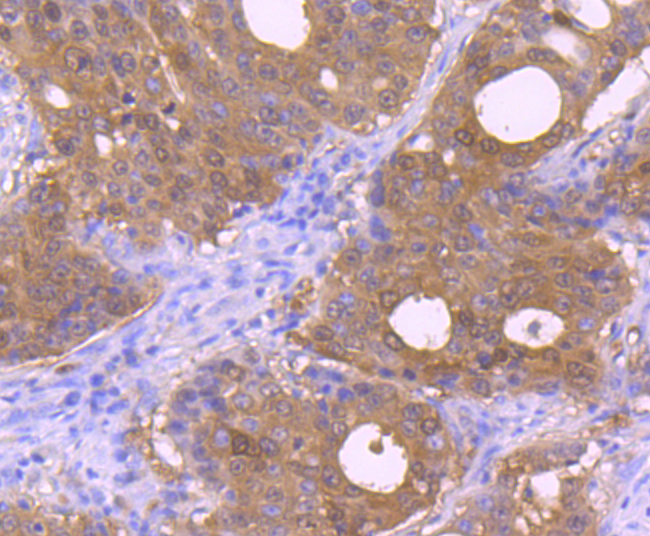
Fig8: Immunohistochemical analysis of paraffin-embedded human stomach cancer tissue using anti-Carbonic anhydrase 2 antibody. Counter stained with hematoxylin.| Product Name | Carbonic anhydrase 2 |
|---|---|
| Antibody Type | Primary Antibodies |
| Antigen Alias | can antibody CAN_ECOLI antibody Carbonate dehydratase 2 antibody Carbonic anhydrase 2 antibody |
| Product description | Carbonic anhydrases (CAs) are members of a large family of zinc metalloenzymes that catalyze the reversible hydration of carbon dioxide. CAs are involved in a variety of biological processes including respiration, calcification, acid-base balance and bone resorption, as well as the formation of aqueous humor, cerebrospinal fluid, saliva and gastric juice. They show extensive diversity in distribution and in their subcellular localization. The human CA2 gene, which maps to chromosome 8q21, encodes CA II, a cytoplasmic protein that has the highest turnover rate and widest tissue distribution of any known human CA isozyme. The human CA4 gene, which maps to chromosome 17q23, encodes CA IV, a membrane-anchored isozyme that is expressed on the luminal surfaces of pulmonary capillaries and proximal renal tubules. The human CA9, CA12 and CA14 genes, which map to chromosomes 9p13, 15q22 and 1q21, respectively, encode transmembrane proteins that have unique patterns of tissue-specific expression. CA IX is specifically expressed in clear-cell renal carcinomas, whereas CA XII is highly expressed in normal tissues, such as kidney, colon and pancreas. Human CA XIV is also expressed in normal tissues, such as brain, but differs from CA XII in its expression pattern. |
| Immunogen | Recombinant protein with human Carbonic anhydrase 2 aa 50-220. |
| Clonality | Polyclonal |
|---|---|
| Isotype | IgG |
| Host Species | Rabbit |
| Tested Applications | WBICCIHC |
| WB:1:500-1:1,000 ICC:1:50-1:200 IHC:1:50-1:200 |
|
| Species Reactivity | Human |
| Concentration | 1mg/ml |
| Alternative Names | can antibody CAN_ECOLI antibody Carbonate dehydratase 2 antibody Carbonic anhydrase 2 antibody |
|---|---|
| Molecular Weight(MW) | 29 kDa |
| Cellular Localization | Cytoplasm. Cell membrane. |
| SwissProt ID | P00918 |
|---|
Application
Fig1: Western blot analysis of Carbonic anhydrase 2 on different cell lysates using anti-Carbonic anhydrase 2 antibody at 1/1,000 dilution. Positive control: Lane1: 293 Lane2: THP-1 Lane3: HL-60
Application
Fig2: ICC staining Carbonic anhydrase 2 in 293T cells (green). The nuclear counter stain is DAPI (blue). Cells were fixed in paraformaldehyde, permeabilised with 0.25% Triton X100/PBS.
Application
Fig3: ICC staining Carbonic anhydrase 2 in Hela cells (green). The nuclear counter stain is DAPI (blue). Cells were fixed in paraformaldehyde, permeabilised with 0.25% Triton X100/PBS.
Application
Fig4: ICC staining Carbonic anhydrase 2 in LOVO cells (green). The nuclear counter stain is DAPI (blue). Cells were fixed in paraformaldehyde, permeabilised with 0.25% Triton X100/PBS.
Application
Fig5: Immunohistochemical analysis of paraffin-embedded human liver tissue using anti-Carbonic anhydrase 2 antibody. Counter stained with hematoxylin.
Application
Fig6: Immunohistochemical analysis of paraffin-embedded human colon cancer tissue using anti-Carbonic anhydrase 2 antibody. Counter stained with hematoxylin.
Application
Fig7: Immunohistochemical analysis of paraffin-embedded human appendix tissue using anti-Carbonic anhydrase 2 antibody. Counter stained with hematoxylin.
Application
Fig8: Immunohistochemical analysis of paraffin-embedded human stomach cancer tissue using anti-Carbonic anhydrase 2 antibody. Counter stained with hematoxylin.| Positive Control | 293, THP-1, HL-60, 293T, Hela, LOVO, human liver tissue, human colon cancer tissue, human appendix tissue, human stomach cancer tissue. |
|---|---|
| Application Notes | WB:1:500-1:1,000 ICC:1:50-1:200 IHC:1:50-1:200 |
| Form | Liquid |
|---|---|
| Storage Instructions | Store at +4℃ after thawing. Aliquot store at -20℃. Avoid repeated freeze / thaw cycles. |
| Storage Buffer | 1*TBS (pH7.4), 0.5%BSA, 50%Glycerol. Preservative: 0.05% Sodium Azide. |


