Application
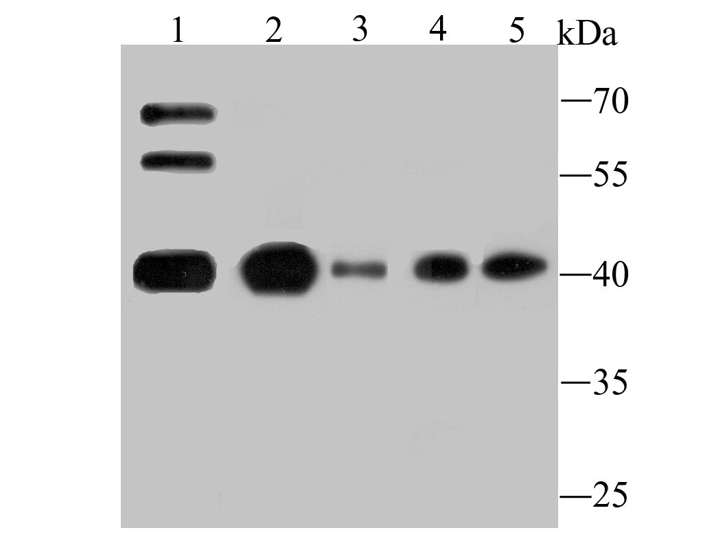
Fig1: Western blot analysis of Apg3 on different lysates using anti-Apg3 antibody at 1/500 dilution. Positive control: Lane 1: Mouse testis tissue Lane 2: K562 Lane 3: HL-60 Lane 4: Hela Lane 5: JurkatApplication
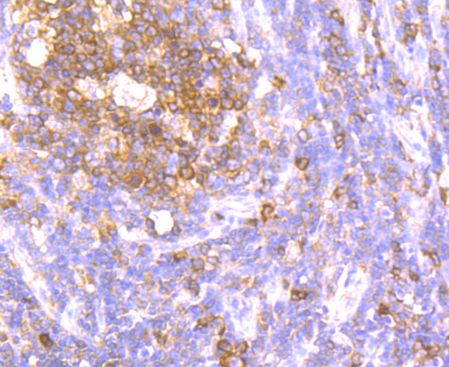
Fig2: Immunohistochemical analysis of paraffin-embedded human tonsil tissue using anti-Apg3 antibody. Counter stained with hematoxylin.Application
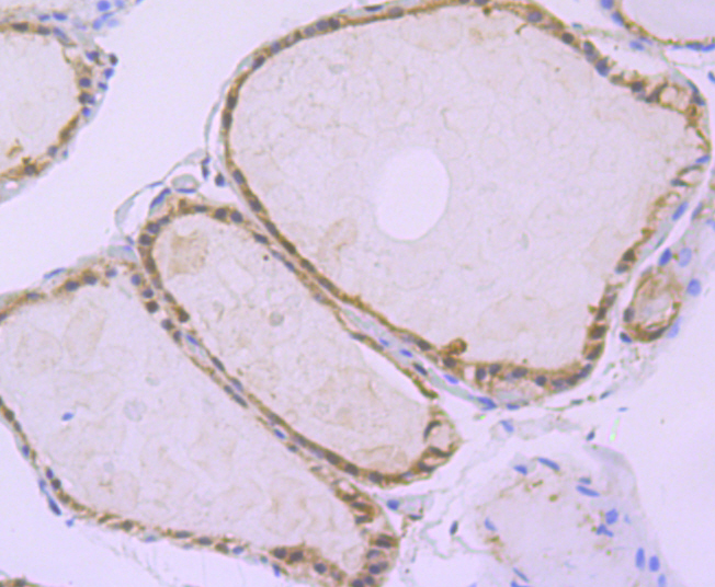
Fig3: Immunohistochemical analysis of paraffin-embedded human thyroid gland tissue using anti-Apg3 antibody. Counter stained with hematoxylin.Application
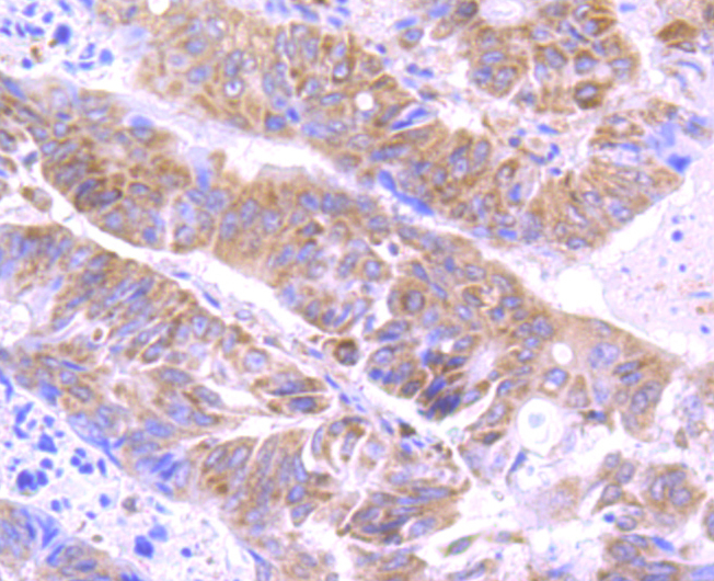
Fig4: Immunohistochemical analysis of paraffin-embedded human colon cancer tissue using anti-Apg3 antibody. Counter stained with hematoxylin.Application
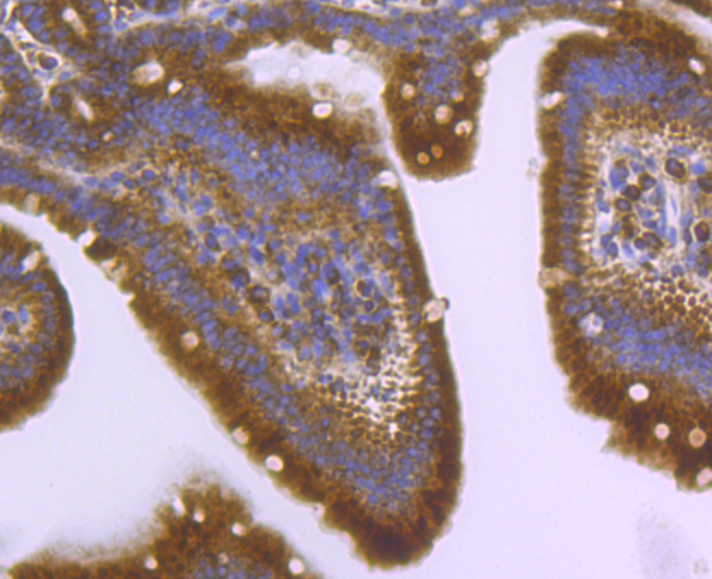
Fig5: Immunohistochemical analysis of paraffin-embedded mouse small intestine tissue using anti-Apg3 antibody. Counter stained with hematoxylin.| Product Name | Apg3 [JU00-35] |
|---|---|
| Antibody Type | Primary Antibodies |
| Antigen Alias | 2610016C12Rik antibody Apg 3 antibody APG3 antibody APG3 autophagy 3 like antibody APG3 like antibody APG3, S. cerevisiae, homolog of antibody APG3-like antibody APG3L antibody Apg3p antibody ATG 3 antibody ATG3 antibody ATG3 autophagy related 3 homolog antibody ATG3 autophagy related 3 homolog (S. cerevisiae) antibody ATG3_HUMAN antibody Autophagy 3, S. cerevisiae, homolog of antibody Autophagy Apg3p/Aut1p like antibody autophagy related 3 antibody Autophagy related protein 3 antibody Autophagy-related protein 3 antibody DKFZp564M1178 antibody FLJ22125 antibody hApg3 antibody MGC15201 antibody OTTHUMP00000214547 antibody OTTHUMP00000214548 antibody PC3 96 antibody Protein PC3-96 antibody Ubiquitin-like-conjugating enzyme ATG3 antibody |
| Product description | Atg3 (autophagy-related protein 3), also known as APG3-like, hAPG3 or PC3-96, is an E2-like enzyme that localizes to the cytoplasm and is expressed in a variety of tissues with predominant levels found in kidney, placenta, liver, heart and skeletal muscle. Atg3 catalyzes the formation of the Atg8-phosphatidylethanolamine (Atg8-PE) conjugate, a reaction that is essential for autophagy (a cellular process that allows for the degradation of organelles and bulk cellular proteins). The process of forming the Atg8-PE conjugate begins with the removal of the C-terminal arginine residue of Atg8 by Atg4, a cysteine protease. The, now exposed, glycine residue is then activated by Atg7 and is then transferred to Atg3 for the final conjugation to PE. This last step can be accelerated by the presence of the Atg12-Atg5 conjugate which functions similarly to an E3 enzyme. |
| Immunogen | Recombinant protein |
| Clonality | Monoclonal |
|---|---|
| Isotype | IgG |
| Host Species | Recombinant rabbit |
| Tested Applications | WBIHC |
| WB:1:500-1:2,000 IHC:1:50-1:200 |
|
| Species Reactivity | HumanMouseRat |
| Concentration | 1mg/ml |
| Alternative Names | 2610016C12Rik antibody
Apg 3 antibody
APG3 antibody
APG3 autophagy 3 like antibody
APG3 like antibody
APG3 S. cerevisiae homolog of antibody APG3-like antibody APG3L antibody Apg3p antibody ATG 3 antibody ATG3 antibody ATG3 autophagy related 3 homolog antibody ATG3 autophagy related 3 homolog (S. cerevisiae) antibody ATG3_HUMAN antibody Autophagy 3 S. cerevisiae homolog of antibody Autophagy Apg3p/Aut1p like antibody autophagy related 3 antibody Autophagy related protein 3 antibody Autophagy-related protein 3 antibody DKFZp564M1178 antibody FLJ22125 antibody hApg3 antibody MGC15201 antibody OTTHUMP00000214547 antibody OTTHUMP00000214548 antibody PC3 96 antibody Protein PC3-96 antibody Ubiquitin-like-conjugating enzyme ATG3 antibody |
|---|---|
| Molecular Weight(MW) | 40 kDa |
| Cellular Localization | Cytoplasm. |
Application
Fig1: Western blot analysis of Apg3 on different lysates using anti-Apg3 antibody at 1/500 dilution. Positive control: Lane 1: Mouse testis tissue Lane 2: K562 Lane 3: HL-60 Lane 4: Hela Lane 5: Jurkat
Application
Fig2: Immunohistochemical analysis of paraffin-embedded human tonsil tissue using anti-Apg3 antibody. Counter stained with hematoxylin.
Application
Fig3: Immunohistochemical analysis of paraffin-embedded human thyroid gland tissue using anti-Apg3 antibody. Counter stained with hematoxylin.
Application
Fig4: Immunohistochemical analysis of paraffin-embedded human colon cancer tissue using anti-Apg3 antibody. Counter stained with hematoxylin.
Application
Fig5: Immunohistochemical analysis of paraffin-embedded mouse small intestine tissue using anti-Apg3 antibody. Counter stained with hematoxylin.| Positive Control | Mouse testis tissue lysate, K562, HL-60, Hela, Jurkat, human tonsil tissue, human thyroid gland tissue, human colon cancer tissue, mouse small intestine tissue. |
|---|---|
| Application Notes | WB:1:500-1:2,000 IHC:1:50-1:200 |
| Form | Liquid |
|---|---|
| Storage Instructions | Store at +4℃ after thawing. Aliquot store at -20℃ or -80℃. Avoid repeated freeze / thaw cycles. |
| Storage Buffer | 1*TBS (pH7.4), 1%BSA, 40%Glycerol. Preservative: 0.05% Sodium Azide. |


