Application
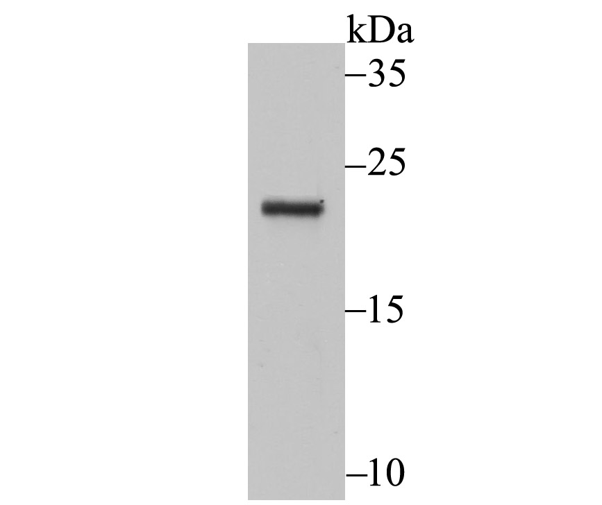
Fig1: Western blot analysis of Securin on HUVEC cell lysate using anti-Securin antibody at 1/500 dilution.Application
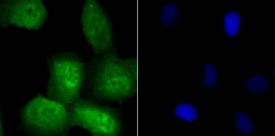
Fig2: ICC staining Securin in HUVEC cells (green). The nuclear counter stain is DAPI (blue). Cells were fixed in paraformaldehyde, permeabilised with 0.25% Triton X100/PBS.Application
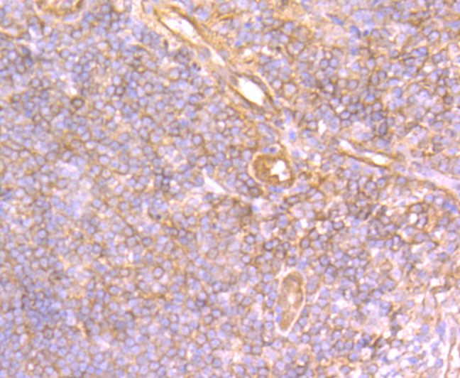
Fig3: Immunohistochemical analysis of paraffin-embedded human tonsil tissue using anti-Securin antibody. Counter stained with hematoxylin.Application
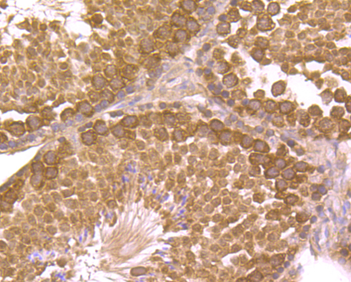
Fig4: Immunohistochemical analysis of paraffin-embedded mouse tetis tissue using anti-Securin antibody. Counter stained with hematoxylin.Application
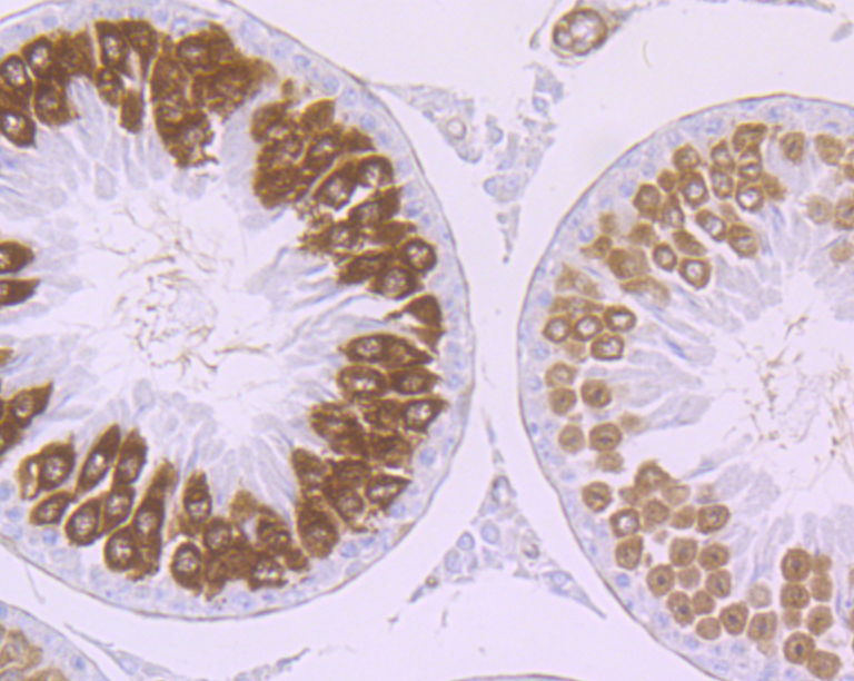
Fig5: Immunohistochemical analysis of paraffin-embedded rat testis tissue using anti-Securin antibody. Counter stained with hematoxylin.Application
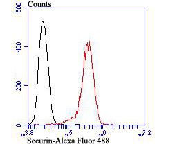
Fig6: Flow cytometric analysis of PC-3M cells with Securin antibody at 1/100 dilution (red) compared with an unlabelled control (cells without incubation with primary antibody; black). Alexa Fluor 488-conjugated goat anti-rabbit IgG was used as the secondary antibody.| Product Name | Securin |
|---|---|
| Antibody Type | Primary Antibodies |
| Antigen Alias | AW555095 antibody C87862 antibody Cut2 antibody EAP 1 antibody EAP1 antibody ESP1 associated protein 1 antibody Esp1-associated protein antibody hPTTG antibody MGC126883 antibody MGC138276 antibody Pds1 antibody Pituitary tumor transforming 1 antibody Pituitary tumor transforming protein 1 antibody Pituitary tumor-transforming 1, isoform CRA_a antibody Pituitary tumor-transforming 1, isoform CRA_b antibody Pituitary tumor-transforming gene 1 antibody Pituitary tumor-transforming gene 1 protein antibody PTTG 1 antibody PTTG antibody PTTG1 antibody PTTG1 protein antibody PTTG1_HUMAN antibody Pttg3 antibody Securin antibody Tumor transforming 1 antibody Tumor transforming protein 1 antibody Tumor-transforming protein 1 antibody TUTR 1 antibody TUTR1 antibody |
| Product description | The product of the oncogene PTTG, pituitary tumor transforming gene, is a human homolog of the anaphase-inhibitor vertebrate protein, securin. PTTG contains a basic amino-terminal domain and an acidic carboxy-terminal domain, which acts as a transactivation domain when fused to a heterologous DNA binding domain. Human PTTG is overexpressed in Jurkat and is also detected in human thymus, testis and placenta. PTTG is mainly expressed in the cytoplasm and is also partially localized to the nucleus. Vertebrate PTTG regulates the separin Esp1, which promotes chromatid separation, to overcome the cohesive forces that hold sister chromatids together. This regulatory function of PTTG suggests that defective regulation of cohesion may contribute to cancer by promoting chromosome instability. Although vertebrate PTTG shares cell-cycle functions with its yeast securin counterparts Pds1p and Cut2, none share sequence homology. |
| Immunogen | Recombinant protein with human Securin aa 1-200. |
| Clonality | Polyclonal |
|---|---|
| Isotype | IgG |
| Host Species | Rabbit |
| Tested Applications | WBICCIHCFC |
| WB:1:500 IHC:1:200-1:500 ICC:1:200-1:500 FC:1:50-1:100 |
|
| Species Reactivity | Human |
| Concentration | 1mg/ml |
| Alternative Names | AW555095 antibody
C87862 antibody
Cut2 antibody
EAP 1 antibody
EAP1 antibody
ESP1 associated protein 1 antibody
Esp1-associated protein antibody
hPTTG antibody
MGC126883 antibody
MGC138276 antibody
Pds1 antibody
Pituitary tumor transforming 1 antibody
Pituitary tumor transforming protein 1 antibody
Pituitary tumor-transforming 1 isoform CRA_a antibody Pituitary tumor-transforming 1 isoform CRA_b antibody Pituitary tumor-transforming gene 1 antibody Pituitary tumor-transforming gene 1 protein antibody PTTG 1 antibody PTTG antibody PTTG1 antibody PTTG1 protein antibody PTTG1_HUMAN antibody Pttg3 antibody Securin antibody Tumor transforming 1 antibody Tumor transforming protein 1 antibody Tumor-transforming protein 1 antibody TUTR 1 antibody TUTR1 antibody |
|---|---|
| Molecular Weight(MW) | 22 kDa |
| Cellular Localization | Nucleus. Cytoplasm. |
| SwissProt ID | O95997 |
|---|
Application
Fig1: Western blot analysis of Securin on HUVEC cell lysate using anti-Securin antibody at 1/500 dilution.
Application
Fig2: ICC staining Securin in HUVEC cells (green). The nuclear counter stain is DAPI (blue). Cells were fixed in paraformaldehyde, permeabilised with 0.25% Triton X100/PBS.
Application
Fig3: Immunohistochemical analysis of paraffin-embedded human tonsil tissue using anti-Securin antibody. Counter stained with hematoxylin.
Application
Fig4: Immunohistochemical analysis of paraffin-embedded mouse tetis tissue using anti-Securin antibody. Counter stained with hematoxylin.
Application
Fig5: Immunohistochemical analysis of paraffin-embedded rat testis tissue using anti-Securin antibody. Counter stained with hematoxylin.
Application
Fig6: Flow cytometric analysis of PC-3M cells with Securin antibody at 1/100 dilution (red) compared with an unlabelled control (cells without incubation with primary antibody; black). Alexa Fluor 488-conjugated goat anti-rabbit IgG was used as the secondary antibody.| Positive Control | HUVEC, PC-3M, human tonsil tissue, mouse testis tissue, rat testis tissue. |
|---|---|
| Application Notes | WB:1:500 IHC:1:200-1:500 ICC:1:200-1:500 FC:1:50-1:100 |
| Form | Liquid |
|---|---|
| Storage Instructions | Store at +4℃ after thawing. Aliquot store at -20℃. Avoid repeated freeze / thaw cycles. |
| Storage Buffer | 1*TBS (pH7.4), 0.5%BSA, 50%Glycerol. Preservative: 0.05% Sodium Azide. |


