Application
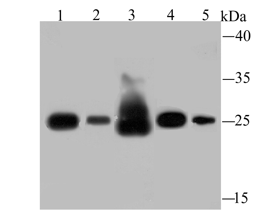
Fig1: Western blot analysis of SDHB on different lysates using anti-SDHB antibody at 1/500 dilution. Positive control: Lane 1: Human liver Lane 2: Rat spleen Lane 3: Rat liver Lane 4: Mouse spleen Lane 5: HepG2Application
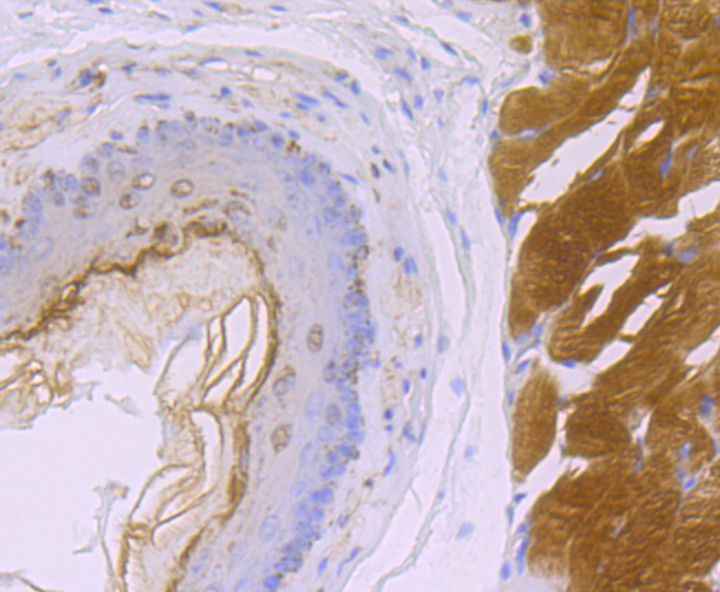
Fig2: Immunohistochemical analysis of paraffin-embedded rat esophagus tissue using anti-SDHB antibody. Counter stained with hematoxylin.Application
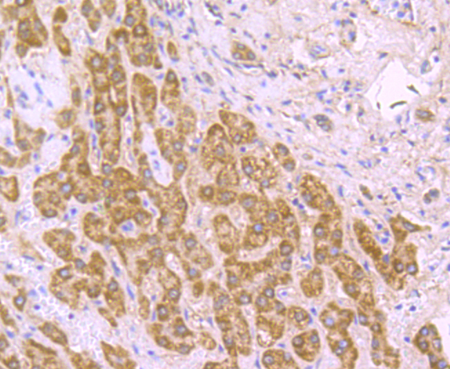
Fig3: Immunohistochemical analysis of paraffin-embedded human liver tissue using anti-SDHB antibody. Counter stained with hematoxylin.Application
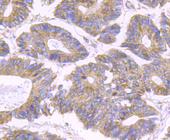
Fig4: Immunohistochemical analysis of paraffin-embedded human colon cancer tissue using anti-SDHB antibody. Counter stained with hematoxylin.Application
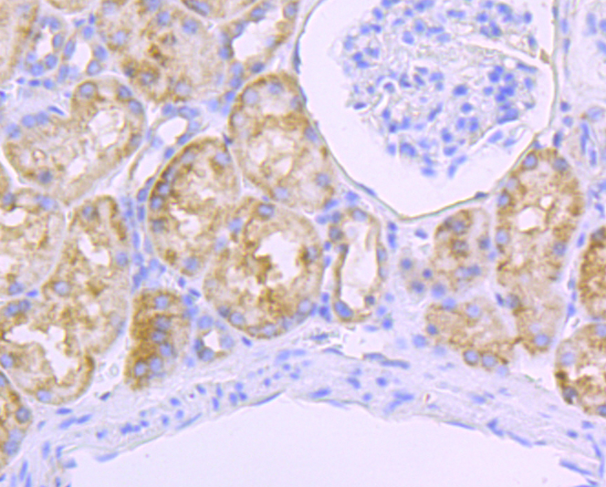
Fig5: Immunohistochemical analysis of paraffin-embedded human kidney tissue using anti-SDHB antibody. Counter stained with hematoxylin.Application
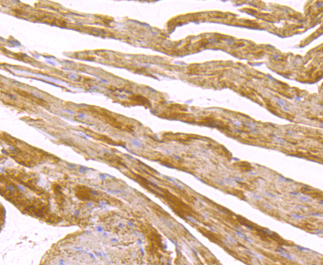
Fig6: Immunohistochemical analysis of paraffin-embedded mouse heart tissue using anti-SDHB antibody. Counter stained with hematoxylin.Application
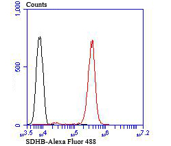
Fig7: Flow cytometric analysis of Jurkat cells with SDHB antibody at 1/100 dilution (red) compared with an unlabelled control (cells without incubation with primary antibody; black). Alexa Fluor® 488-conjugated goat anti-rabbit IgG was used as the secondary antibody.| Product Name | SDHB [JU02-33] |
|---|---|
| Antibody Type | Primary Antibodies |
| Antigen Alias | CWS2 antibody DHSB_HUMAN antibody FLJ92337 antibody Ip antibody Iron sulfur subunit antibody Iron sulfur subunit of complex II antibody Iron-sulfur subunit of complex II antibody mitochondrial antibody PGL 4 antibody PGL4 antibody SDH 1 antibody SDH antibody SDH1 antibody SDH2 antibody SDH2, homolog of antibody SdhB antibody SDHIP antibody Succinate dehydrogenase [ubiquinone] iron sulfur protein mitochondrial antibody Succinate dehydrogenase [ubiquinone] iron sulfur subunit antibody Succinate dehydrogenase [ubiquinone] iron-sulfur subunit antibody succinate dehydrogenase [ubiquinone] iron-sulfur subunit, mitochondrial antibody Succinate Dehydrogenase 1 Iron Sulfur Subunit antibody Succinate Dehydrogenase 2, S. cerevisiae, homolog of antibody Succinate dehydrogenase complex iron sulfur subunit B antibody Succinate dehydrogenase complex subunit B iron sulfur antibody Succinate Dehydrogenase Complex Subunit B Iron Sulfur Protein antibody succinate dehydrogenase complex, subunit B, iron sulfur (Ip) antibody Succinate dehydrogenase iron sulfur protein antibody |
| Product description | In aerobic respiration reactions, succinate dehydrogenase (SDH) catalyzes the oxidation of succinate and ubiquinone to fumarate and ubiquinol. Four subunits comprise the SDH protein complex: a flavochrome subunit (SDHA), an iron-sulfur protein (SDHB) and two membrane-bound subunits (SDHC and SDHD) anchored to the inner mitochondrial membrane. Mutations to these subunits cause mitochondrial dysfunction, corresponding to several distinct disorders. Mutations in the membrane bound components may cause hereditary paraganglioma, while SDHA mutations associate with juvenile encephalopathy as well as Leigh syndrome, a severe neurological disorder. Inactivating mutations in SDHB correlate with inherited, but not necessarily sporadic, cases of pheochromocytoma. |
| Immunogen | Recombinant protein |
| Clonality | Monoclonal |
|---|---|
| Isotype | IgG |
| Host Species | Recombinant rabbit |
| Tested Applications | WBIHCFCIP |
| WB:1:500-1:2,000 IHC:1:50-1:200 FC:1:50-1:100 IP:1:10-1:50 |
|
| Species Reactivity | HumanMouseRat |
| Concentration | 1mg/ml |
| Alternative Names | CWS2 antibody
DHSB_HUMAN antibody
FLJ92337 antibody Ip antibody
Iron sulfur subunit antibody
Iron sulfur subunit of complex II antibody
Iron-sulfur subunit of complex II antibody
mitochondrial antibody
PGL 4 antibody
PGL4 antibody
SDH 1 antibody
SDH antibody
SDH1 antibody
SDH2 antibody
SDH2 homolog of antibody SdhB antibody SDHIP antibody Succinate dehydrogenase [ubiquinone] iron sulfur protein mitochondrial antibody Succinate dehydrogenase [ubiquinone] iron sulfur subunit antibody Succinate dehydrogenase [ubiquinone] iron-sulfur subunit antibody succinate dehydrogenase [ubiquinone] iron-sulfur subunit mitochondrial antibody Succinate Dehydrogenase 1 Iron Sulfur Subunit antibody Succinate Dehydrogenase 2 S. cerevisiae homolog of antibody Succinate dehydrogenase complex iron sulfur subunit B antibody Succinate dehydrogenase complex subunit B iron sulfur antibody Succinate Dehydrogenase Complex Subunit B Iron Sulfur Protein antibody succinate dehydrogenase complex subunit B iron sulfur (Ip) antibody Succinate dehydrogenase iron sulfur protein antibody |
|---|---|
| Molecular Weight(MW) | 32 kDa |
| Cellular Localization | Mitochondrion. Mitochondrion inner membrane. |
Application
Fig1: Western blot analysis of SDHB on different lysates using anti-SDHB antibody at 1/500 dilution. Positive control: Lane 1: Human liver Lane 2: Rat spleen Lane 3: Rat liver Lane 4: Mouse spleen Lane 5: HepG2
Application
Fig2: Immunohistochemical analysis of paraffin-embedded rat esophagus tissue using anti-SDHB antibody. Counter stained with hematoxylin.
Application
Fig3: Immunohistochemical analysis of paraffin-embedded human liver tissue using anti-SDHB antibody. Counter stained with hematoxylin.
Application
Fig4: Immunohistochemical analysis of paraffin-embedded human colon cancer tissue using anti-SDHB antibody. Counter stained with hematoxylin.
Application
Fig5: Immunohistochemical analysis of paraffin-embedded human kidney tissue using anti-SDHB antibody. Counter stained with hematoxylin.
Application
Fig6: Immunohistochemical analysis of paraffin-embedded mouse heart tissue using anti-SDHB antibody. Counter stained with hematoxylin.
Application
Fig7: Flow cytometric analysis of Jurkat cells with SDHB antibody at 1/100 dilution (red) compared with an unlabelled control (cells without incubation with primary antibody; black). Alexa Fluor® 488-conjugated goat anti-rabbit IgG was used as the secondary antibody.| Positive Control | Human liver tissue, rat spleen tissue lysate, rat liver tissue lysate, mouse spleen tissue lysate, HepG2, rat esophagus tissue, human colon cancer tissue, human kidney tissue, mouse heart tissue, Jurkat. |
|---|---|
| Application Notes | WB:1:500-1:2,000 IHC:1:50-1:200 FC:1:50-1:100 IP:1:10-1:50 |
| Form | Liquid |
|---|---|
| Storage Instructions | Store at +4℃ after thawing. Aliquot store at -20℃ or -80℃. Avoid repeated freeze / thaw cycles. |
| Storage Buffer | 1*TBS (pH7.4), 1%BSA, 40%Glycerol. Preservative: 0.05% Sodium Azide. |


