Application
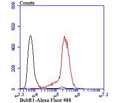
Fig6: Flow cytometric analysis of HL-60 cells with BubR1 antibody at 1/100 dilution (red) compared with an unlabelled control (cells without incubation with primary antibody; black). Alexa Fluor 488-conjugated goat anti-rabbit IgG was used as the secondary antibody.Application
Fig6: Flow cytometric analysis of HL-60 cells with BubR1 antibody at 1/100 dilution (red) compared with an unlabelled control (cells without incubation with primary antibody; black). Alexa Fluor 488-conjugated goat anti-rabbit IgG was used as the secondary antibody.Application
Fig6: Flow cytometric analysis of HL-60 cells with BubR1 antibody at 1/100 dilution (red) compared with an unlabelled control (cells without incubation with primary antibody; black). Alexa Fluor 488-conjugated goat anti-rabbit IgG was used as the secondary antibody.Application
Fig6: Flow cytometric analysis of HL-60 cells with BubR1 antibody at 1/100 dilution (red) compared with an unlabelled control (cells without incubation with primary antibody; black). Alexa Fluor 488-conjugated goat anti-rabbit IgG was used as the secondary antibody.Application
Fig6: Flow cytometric analysis of HL-60 cells with BubR1 antibody at 1/100 dilution (red) compared with an unlabelled control (cells without incubation with primary antibody; black). Alexa Fluor 488-conjugated goat anti-rabbit IgG was used as the secondary antibody.Application
Fig6: Flow cytometric analysis of HL-60 cells with BubR1 antibody at 1/100 dilution (red) compared with an unlabelled control (cells without incubation with primary antibody; black). Alexa Fluor 488-conjugated goat anti-rabbit IgG was used as the secondary antibody.| Product Name | BubR1 |
|---|---|
| Antibody Type | Primary Antibodies |
| Antigen Alias | Beta homolg of S. cerevisiae BUB 1 antibody Beta homolg of S. cerevisiae budding uninhibited by benzimidazoles antibody BUB 1B antibody BUB1 budding uninhibited by benzimidazoles 1 homolog beta antibody Bub1A antibody BUB1B antibody BUB1B_HUMAN antibody BUB1beta antibody BUBR1 antibody Budding Uninhibited by Benzimidazoles 1 beta antibody Budding uninhibited by benzimidazoles 1 homolog beta (yeast) antibody hBUBR1 antibody MAD3/BUB1 related protein kinase antibody MAD3/BUB1-related protein kinase antibody MAD3L antibody Mitotic checkpoint gene BUB1B antibody Mitotic checkpoint kinase MAD3L antibody Mitotic checkpoint serine/threonine protein kinase BUB1 beta antibody Mitotic checkpoint serine/threonine-protein kinase BUB1 beta antibody MVA1 antibody OTTHUMP00000160319 antibody Protein SSK1 antibody SSK 1 antibody SSK1 antibody |
| Product description | Human cells contain two related protein kinases, BUB1 and BUBR1, that appear to have evolved from a single ancestral BUB1 gene. Both kinases are concentrated near the surface of the kinetochore where they monitor kinetochore-microtubule interactions. BUB1 and BUBR1 bind to kinetochores and are postulated to be components of the mitotic checkpoint, which monitors kinetochore activities to determine if chromosomes have achieved alignment at the spindle equator. BUBR1 is essential for normal mitotic progression as it prevents cells from prematurely entering anaphase. BUB3 is a conserved component of the mitotic spindle assembly complex and is also involved with the essential spindle checkpoint pathway that operates during early embryogenesis. |
| Immunogen | Recombinant protein within human BubR1 330-600 aa. |
| Clonality | Polyclonal |
|---|---|
| Isotype | IgG |
| Host Species | Rabbit |
| Tested Applications | WBICCIHCFC |
| WB:1:5,000-1:10,000 ICC:1:50-1:200 IHC:1:50-1:200 FC:1:50-1:100 |
|
| Species Reactivity | Human |
| Concentration | 1mg/ml |
| Alternative Names | Beta homolg of S. cerevisiae BUB 1 antibody Beta homolg of S. cerevisiae budding uninhibited by benzimidazoles antibody BUB 1B antibody BUB1 budding uninhibited by benzimidazoles 1 homolog beta antibody Bub1A antibody BUB1B antibody BUB1B_HUMAN antibody BUB1beta antibody BUBR1 antibody Budding Uninhibited by Benzimidazoles 1 beta antibody Budding uninhibited by benzimidazoles 1 homolog beta (yeast) antibody hBUBR1 antibody MAD3/BUB1 related protein kinase antibody MAD3/BUB1-related protein kinase antibody MAD3L antibody Mitotic checkpoint gene BUB1B antibody Mitotic checkpoint kinase MAD3L antibody Mitotic checkpoint serine/threonine protein kinase BUB1 beta antibody Mitotic checkpoint serine/threonine-protein kinase BUB1 beta antibody MVA1 antibody OTTHUMP00000160319 antibody Protein SSK1 antibody SSK 1 antibody SSK1 antibody |
|---|---|
| Molecular Weight(MW) | 119 kDa |
| Cellular Localization | Nucleus. Cytoplasm. |
| SwissProt ID | O60566 |
|---|
Application
Fig6: Flow cytometric analysis of HL-60 cells with BubR1 antibody at 1/100 dilution (red) compared with an unlabelled control (cells without incubation with primary antibody; black). Alexa Fluor 488-conjugated goat anti-rabbit IgG was used as the secondary antibody.
Application
Fig6: Flow cytometric analysis of HL-60 cells with BubR1 antibody at 1/100 dilution (red) compared with an unlabelled control (cells without incubation with primary antibody; black). Alexa Fluor 488-conjugated goat anti-rabbit IgG was used as the secondary antibody.
Application
Fig6: Flow cytometric analysis of HL-60 cells with BubR1 antibody at 1/100 dilution (red) compared with an unlabelled control (cells without incubation with primary antibody; black). Alexa Fluor 488-conjugated goat anti-rabbit IgG was used as the secondary antibody.
Application
Fig6: Flow cytometric analysis of HL-60 cells with BubR1 antibody at 1/100 dilution (red) compared with an unlabelled control (cells without incubation with primary antibody; black). Alexa Fluor 488-conjugated goat anti-rabbit IgG was used as the secondary antibody.
Application
Fig6: Flow cytometric analysis of HL-60 cells with BubR1 antibody at 1/100 dilution (red) compared with an unlabelled control (cells without incubation with primary antibody; black). Alexa Fluor 488-conjugated goat anti-rabbit IgG was used as the secondary antibody.
Application
Fig6: Flow cytometric analysis of HL-60 cells with BubR1 antibody at 1/100 dilution (red) compared with an unlabelled control (cells without incubation with primary antibody; black). Alexa Fluor 488-conjugated goat anti-rabbit IgG was used as the secondary antibody.| Positive Control | Hela, SH-SY-5Y, SiHa, human spleen tissue, HL-60. |
|---|---|
| Application Notes | WB:1:5,000-1:10,000 ICC:1:50-1:200 IHC:1:50-1:200 FC:1:50-1:100 |
| Form | Liquid |
|---|---|
| Storage Instructions | Store at +4℃ after thawing. Aliquot store at -20℃. Avoid repeated freeze / thaw cycles. |
| Storage Buffer | 1*TBS (pH7.4), 0.5%BSA, 50%Glycerol. Preservative: 0.05% Sodium Azide. |


