Application
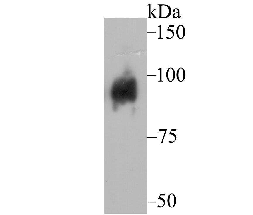
Fig1: Western blot analysis of Dynamin 1 on mouse brain tissue lysate using anti-Dynamin 1 antibody at 1/500 dilution.Application
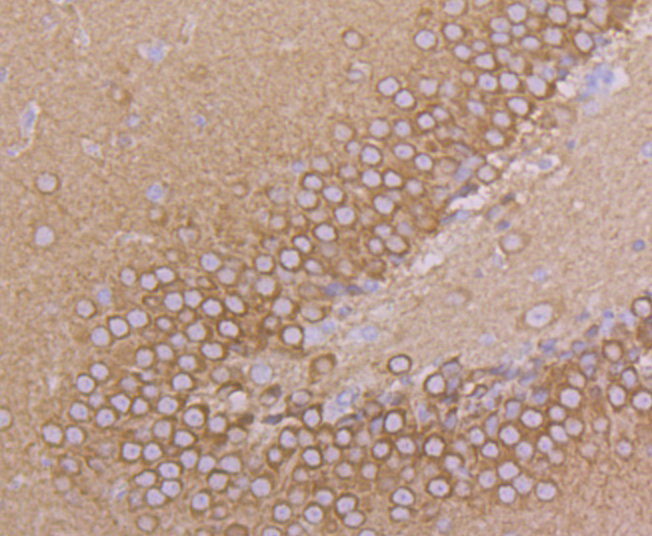
Fig2: Immunohistochemical analysis of paraffin-embedded rat brain tissue using anti-Dynamin 1 antibody. Counter stained with hematoxylin.Application
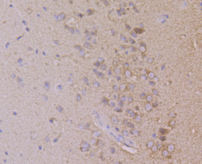
Fig3: Immunohistochemical analysis of paraffin-embedded mouse brain tissue using anti-Dynamin 1 antibody. Counter stained with hematoxylin.Application
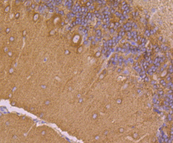
Fig4: Immunohistochemical analysis of paraffin-embedded mouse cerebellum tissue using anti-Dynamin 1 antibody. Counter stained with hematoxylin.| Product Name | Dynamin 1 |
|---|---|
| Antibody Type | Primary Antibodies |
| Antigen Alias | B dynamin antibody D100 antibody DNM 1 antibody DNM antibody DNM1 antibody DYN1_HUMAN antibody Dynamin antibody Dynamin-1 antibody Dynamin1 antibody |
| Product description | Members of the Dynamin family, including Dynamin I and Dynamin II, are GTPase, microtubule-associated proteins which are involved in endocytosis, synaptic transmission and neurogenesis. Dynamin I is localized to the central nervous system, while Dynamin II exhibits ubiquitous distribution with highest expression found in testis. Both dynamin proteins contain SH3 and proline-rich domains that mediate interactions between the dynamins and effectors of their GTPase activity). The interactions with these effectors, which include microtubules, acidic phospholipids and SH3 domain-containing proteins, are required for rapid endocytosis. Dynamin I appears to be recruited to clathrin coated pits by SH3 domain interaction with amphiphysin, a protein highly expressed in brain. |
| Immunogen | Recombinant protein within human Dynamin 1 aa 575-750. |
| Clonality | Polyclonal |
|---|---|
| Isotype | IgG |
| Host Species | Rabbit |
| Tested Applications | WBIHC |
| WB:1:500-1:1,000 IHC:1:50-1:200 |
|
| Species Reactivity | HumanMouseRat |
| Concentration | 1mg/ml |
| Alternative Names | B dynamin antibody D100 antibody DNM 1 antibody DNM antibody DNM1 antibody DYN1_HUMAN antibody Dynamin antibody Dynamin-1 antibody Dynamin1 antibody |
|---|---|
| Molecular Weight(MW) | 97 kDa |
| Cellular Localization | Cytoplasm. Cytoskeleton. |
Application
Fig1: Western blot analysis of Dynamin 1 on mouse brain tissue lysate using anti-Dynamin 1 antibody at 1/500 dilution.
Application
Fig2: Immunohistochemical analysis of paraffin-embedded rat brain tissue using anti-Dynamin 1 antibody. Counter stained with hematoxylin.
Application
Fig3: Immunohistochemical analysis of paraffin-embedded mouse brain tissue using anti-Dynamin 1 antibody. Counter stained with hematoxylin.
Application
Fig4: Immunohistochemical analysis of paraffin-embedded mouse cerebellum tissue using anti-Dynamin 1 antibody. Counter stained with hematoxylin.| Positive Control | Mouse brain tissue, rat brain tissue, mouse cerebellum tissue. |
|---|---|
| Application Notes | WB:1:500-1:1,000 IHC:1:50-1:200 |
| Form | Liquid |
|---|---|
| Storage Instructions | Store at +4℃ after thawing. Aliquot store at -20℃. Avoid repeated freeze / thaw cycles. |
| Storage Buffer | 1*TBS (pH7.4), 0.5%BSA, 50%Glycerol. Preservative: 0.05% Sodium Azide. |


