Application
Fig1: Western blot analysis of BHLHB9 on U937 and SH-SY-5Y cell lysates using anti-BHLHB9 antibody at 1/500 dilution.Application
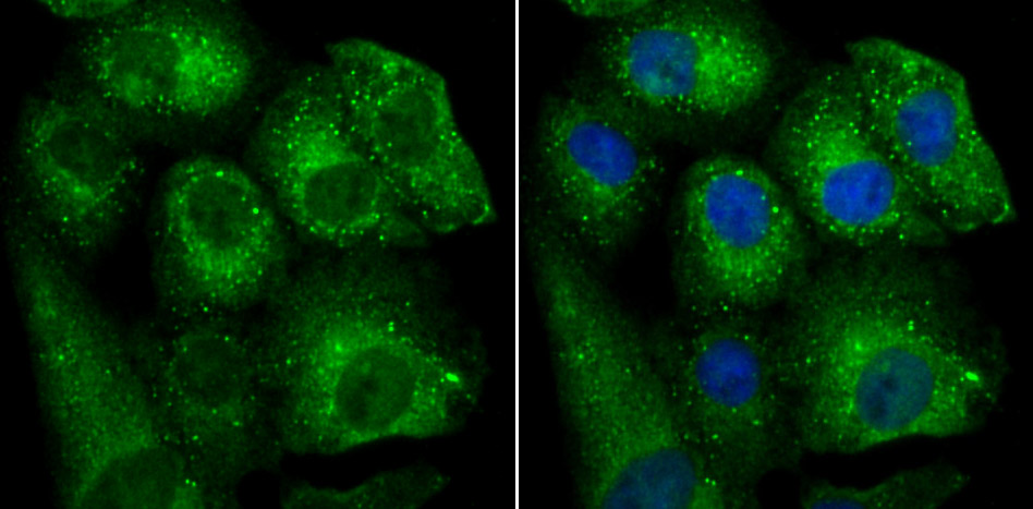
Fig2: ICC staining BHLHB9 in A549 cells (green). The nuclear counter stain is DAPI (blue). Cells were fixed in paraformaldehyde, permeabilised with 0.25% Triton X100/PBS.Application
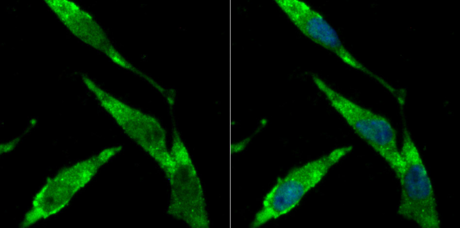
Fig3: ICC staining BHLHB9 in SH-SY-5Y cells (green). The nuclear counter stain is DAPI (blue). Cells were fixed in paraformaldehyde, permeabilised with 0.25% Triton X100/PBS.Application
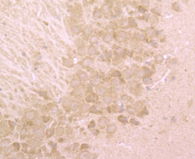
Fig4: Immunohistochemical analysis of paraffin-embedded mouse brain tissue using anti-BHLHB9 antibody. Counter stained with hematoxylin.| Product Name | BHLHB9 |
|---|---|
| Antibody Type | Primary Antibodies |
| Product description | This gene is a member of a gene family which encodes proteins with a basic helix-loop-helix domain. Other members of this gene family encode proteins which function as transcription factors, either enhancing or inhibiting transcription depending on the activity of other DNA binding proteins. The coding region of this gene is located entirely within the terminal exon. The encoded protein may be involved in the survival of neurons. Multiple alternatively spliced variants, encoding the same protein, have been identified. May play a role in the control of cellular aging and survival. Highly expressed in brain. Not expressed in lung or liver. Down-regulated in brain from patients suffering from Alzheimer disease. |
| Immunogen | Peptide |
| Clonality | Polyclonal |
|---|---|
| Isotype | IgG |
| Host Species | Rabbit |
| Tested Applications | WBICCIHC |
| WB:1:500 ICC:1:50-1:200 IHC:1:50-1:200 |
|
| Species Reactivity | HumanMouse |
| Concentration | 1mg/ml |
| Molecular Weight(MW) | 60 kDa |
|---|---|
| Cellular Localization | Nucleus. Cytoplasm. |
Application
Fig1: Western blot analysis of BHLHB9 on U937 and SH-SY-5Y cell lysates using anti-BHLHB9 antibody at 1/500 dilution.
Application
Fig2: ICC staining BHLHB9 in A549 cells (green). The nuclear counter stain is DAPI (blue). Cells were fixed in paraformaldehyde, permeabilised with 0.25% Triton X100/PBS.
Application
Fig3: ICC staining BHLHB9 in SH-SY-5Y cells (green). The nuclear counter stain is DAPI (blue). Cells were fixed in paraformaldehyde, permeabilised with 0.25% Triton X100/PBS.
Application
Fig4: Immunohistochemical analysis of paraffin-embedded mouse brain tissue using anti-BHLHB9 antibody. Counter stained with hematoxylin.| Positive Control | U937, A549, SH-SY-5Y, mouse brain tissue. |
|---|---|
| Application Notes | WB:1:500 ICC:1:50-1:200 IHC:1:50-1:200 |
| Form | Liquid |
|---|---|
| Storage Instructions | Store at +4℃ after thawing. Aliquot store at -20℃ or -80℃. Avoid repeated freeze / thaw cycles. |
| Storage Buffer | 1*TBS (pH7.4), 0.5%BSA, 50%Glycerol. Preservative: 0.05% Sodium Azide. |


