Application
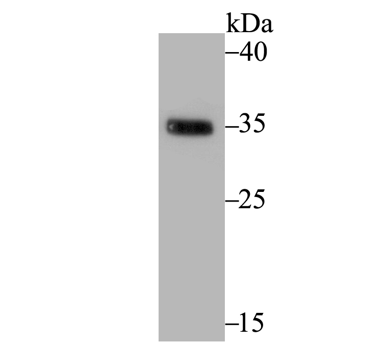
Fig1: Western blot analysis of EpCAM on mouse colon tissue lysate using anti-EpCAM antibody at 1/500 dilution.Application
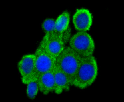
Fig2: ICC staining EpCAM in LOVO cells (green). The nuclear counter stain is DAPI (blue). Cells were fixed in paraformaldehyde, permeabilised with 0.25% Triton X100/PBS.Application
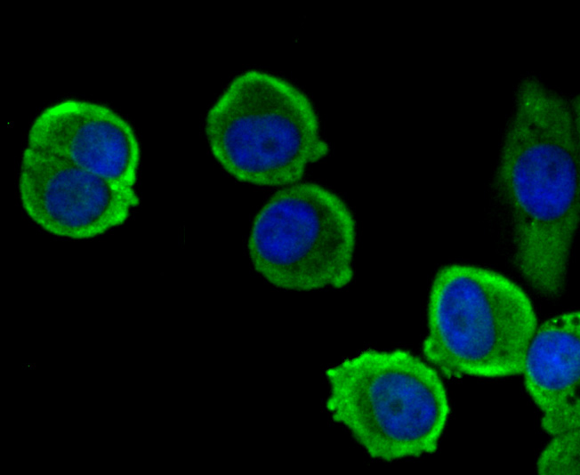
Fig3: ICC staining EpCAM in MCF-7 cells (green). The nuclear counter stain is DAPI (blue). Cells were fixed in paraformaldehyde, permeabilised with 0.25% Triton X100/PBS.Application
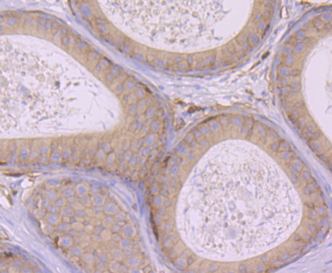
Fig4: Immunohistochemical analysis of paraffin-embedded rat epididymis tissue using anti-EpCAM antibody. Counter stained with hematoxylin.Application
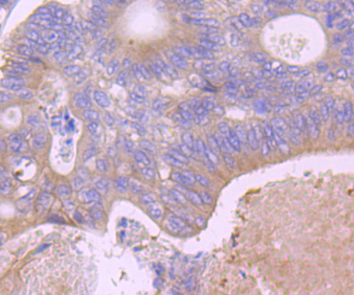
Fig5: Immunohistochemical analysis of paraffin-embedded human colon cancer tissue using anti-EpCAM antibody. Counter stained with hematoxylin.Application
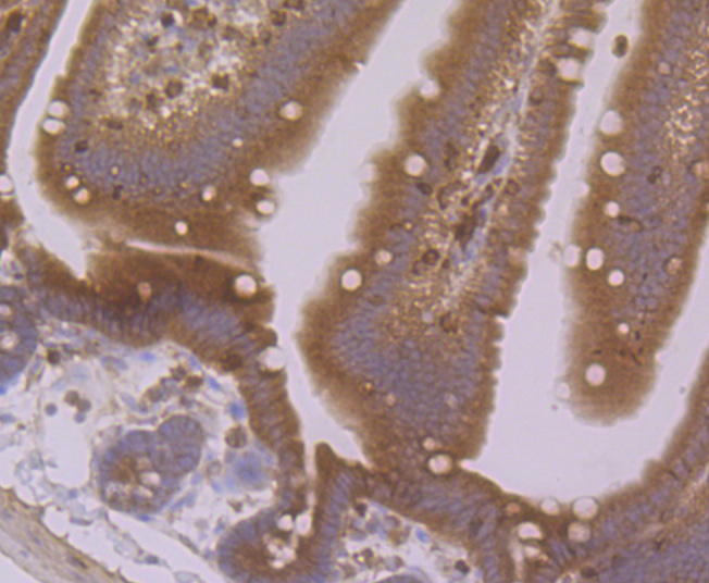
Fig6: Immunohistochemical analysis of paraffin-embedded mouse small intestine tissue using anti-EpCAM antibody. Counter stained with hematoxylin.| Product Name | EpCAM |
|---|---|
| Antibody Type | Primary Antibodies |
| Antigen Alias | 17 1A antibody 323/A3 antibody Adenocarcinoma associated antigen antibody Adenocarcinoma-associated antigen antibody Antigen identified by monoclonal antibody AUA1 antibody AUA1 antibody CD326 antibody CD326 antigen antibody Cell surface glycoprotein Trop 1 antibody Cell surface glycoprotein Trop 2 antibody Cell surface glycoprotein Trop-1 antibody CO 17A antibody CO17 1A antibody CO17A antibody DIAR5 antibody EGP 2 antibody EGP antibody EGP2 antibody EGP314 antibody EGP40 antibody Ep CAM antibody Ep-CAM antibody EPCAM antibody EPCAM_HUMAN antibody EpCAM1 antibody Epithelial cell adhesion molecule antibody Epithelial Cell Adhesion Molecule Intracellular Domain (EpCAM-ICD) antibody Epithelial cell surface antigen antibody Epithelial cellular adhesion molecule antibody Epithelial glycoprotein 1 antibody Epithelial glycoprotein 314 antibody Epithelial glycoprotein antibody ESA antibody GA733 1 antibody GA733 2 antibody GA733-2 antibody gastrointestinal tumor-associated antigen 2, 35-KD glycoprotein antibody gp4 antibody hEGP 2 antibody hEGP314 antibody HNPCC8 antibody Human epithelial glycoprotein 2 antibody KS 1/4 antigen antibody KS1/4 antibody KSA antibody Ly74 antibody Lymphocyte antigen 74 antibody M1S 1 antibody M1S2 antibody M4S1 antibody Major gastrointestinal tumor associated protein GA733 2 antibody Major gastrointestinal tumor-associated protein GA733-2 antibody mEGP314 antibody Membrane component chromosome 4 surface marker (35kD glycoprotein) antibody Membrane component, chromosome 4, surface marker 1 antibody Membrane component, chromosome 4, surface marker antibody MIC18 antibody MK 1 antibody Protein 289A antibody TACD1 antibody TACSTD1 antibody TROP1 antibody Tumor associated calcium signal transducer 1 antibody Tumor associated calcium signal transducer 2 precursor antibody Tumor-associated calcium signal transducer 1 antibody |
| Product description | The epithelial cell adhesion molecule Ep-CAM, which is also designated tumor-associated calcium signal transducer 1 and MK-1, is a monomeric membrane glycoprotein that is expressed in most normal human epithelium and carcinomas. The human Ep-CAM gene encodes a 314 amino acid protein that is expressed as two forms, a major form and a minor form, which are reduced upon treatment with the amino-glycosylation inhibitor tunicamycin. Ep-CAM is overexpressed in a variety of carcinomas and is, therefore, a potential target for the visualization and therapy of human solid tumors. Ep-CAM contains an extracellular domain containing two epidermal growth factor-like repeats, followed by a cysteine poor region, which is necessary for the adhesion properties of the molecule. |
| Immunogen | Synthetic peptide within human EpCAM 100-150 aa. |
| Clonality | Polyclonal |
|---|---|
| Isotype | IgG |
| Host Species | Rabbit |
| Tested Applications | WBICCIHC |
| WB:1:500 ICC:1:100 IHC:1:50-1:200 |
|
| Species Reactivity | HumanMouseRat |
| Concentration | 1mg/ml |
| Alternative Names | 17 1A antibody
323/A3 antibody
Adenocarcinoma associated antigen antibody
Adenocarcinoma-associated antigen antibody
Antigen identified by monoclonal antibody AUA1 antibody
AUA1 antibody
CD326 antibody
CD326 antigen antibody
Cell surface glycoprotein Trop 1 antibody
Cell surface glycoprotein Trop 2 antibody
Cell surface glycoprotein Trop-1 antibody
CO 17A antibody
CO17 1A antibody
CO17A antibody
DIAR5 antibody
EGP 2 antibody
EGP antibody
EGP2 antibody
EGP314 antibody
EGP40 antibody
Ep CAM antibody
Ep-CAM antibody
EPCAM antibody
EPCAM_HUMAN antibody
EpCAM1 antibody
Epithelial cell adhesion molecule antibody
Epithelial Cell Adhesion Molecule Intracellular Domain (EpCAM-ICD) antibody
Epithelial cell surface antigen antibody
Epithelial cellular adhesion molecule antibody
Epithelial glycoprotein 1 antibody
Epithelial glycoprotein 314 antibody
Epithelial glycoprotein antibody
ESA antibody
GA733 1 antibody
GA733 2 antibody
GA733-2 antibody
gastrointestinal tumor-associated antigen 2 35-KD glycoprotein antibody gp4 antibody hEGP 2 antibody hEGP314 antibody HNPCC8 antibody Human epithelial glycoprotein 2 antibody KS 1/4 antigen antibody KS1/4 antibody KSA antibody Ly74 antibody Lymphocyte antigen 74 antibody M1S 1 antibody M1S2 antibody M4S1 antibody Major gastrointestinal tumor associated protein GA733 2 antibody Major gastrointestinal tumor-associated protein GA733-2 antibody mEGP314 antibody Membrane component chromosome 4 surface marker (35kD glycoprotein) antibody Membrane component chromosome 4 surface marker 1 antibody Membrane component chromosome 4 surface marker antibody MIC18 antibody MK 1 antibody Protein 289A antibody TACD1 antibody TACSTD1 antibody TROP1 antibody Tumor associated calcium signal transducer 1 antibody Tumor associated calcium signal transducer 2 precursor antibody Tumor-associated calcium signal transducer 1 antibody |
|---|---|
| Molecular Weight(MW) | 35 kDa |
| Cellular Localization | Cell junction. Plasma membrane. |
Application
Fig1: Western blot analysis of EpCAM on mouse colon tissue lysate using anti-EpCAM antibody at 1/500 dilution.
Application
Fig2: ICC staining EpCAM in LOVO cells (green). The nuclear counter stain is DAPI (blue). Cells were fixed in paraformaldehyde, permeabilised with 0.25% Triton X100/PBS.
Application
Fig3: ICC staining EpCAM in MCF-7 cells (green). The nuclear counter stain is DAPI (blue). Cells were fixed in paraformaldehyde, permeabilised with 0.25% Triton X100/PBS.
Application
Fig4: Immunohistochemical analysis of paraffin-embedded rat epididymis tissue using anti-EpCAM antibody. Counter stained with hematoxylin.
Application
Fig5: Immunohistochemical analysis of paraffin-embedded human colon cancer tissue using anti-EpCAM antibody. Counter stained with hematoxylin.
Application
Fig6: Immunohistochemical analysis of paraffin-embedded mouse small intestine tissue using anti-EpCAM antibody. Counter stained with hematoxylin.| Positive Control | Mouse colon tissue lysate, LOVO, MCF-7, rat epididymis tissue, human colon cancer tissue, mouse small intestine tissue. |
|---|---|
| Application Notes | WB:1:500 ICC:1:100 IHC:1:50-1:200 |
| Form | Liquid |
|---|---|
| Storage Instructions | Store at +4℃ after thawing. Aliquot store at -20℃. Avoid repeated freeze / thaw cycles. |
| Storage Buffer | 1*TBS (pH7.4), 0.5%BSA, 50%Glycerol. Preservative: 0.05% Sodium Azide. |


