Application
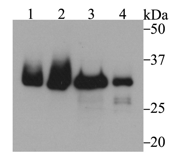
Fig1: Western blot analysis of Fibrillarin on different cell lysates using anti-Fibrillarin antibody at 1/1,000 dilution. Positive control: Lane1: Hela Lane2: HepG2 Lane3: 293T Lane4: NIH-3T3Application
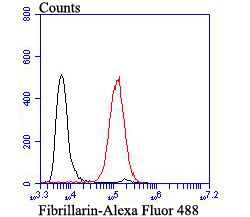
Fig2: Flow cytometric analysis of HL-60 cells with Fibrillarin antibody at 1/100 dilution (red) compared with an unlabelled control (cells without incubation with primary antibody; black). Alexa Fluor 488-conjugated goat anti-rabbit IgG was used as the secondary antibody.| Product Name | Fibrillarin |
|---|---|
| Antibody Type | Primary Antibodies |
| Antigen Alias | 34 kD nucleolar scleroderma antigen antibody 34 kDa nucleolar scleroderma antigen antibody Fbl antibody FBRL_HUMAN antibody FIB antibody FIB1 antibody FLRN antibody Histone-glutamine methyltransferase antibody Nop1p antibody RNA U3 small nucleolar interacting protein 1 antibody RNU3IP1 antibody rRNA 2' O methyltransferase fibrillarin antibody rRNA 2'-O-methyltransferase fibrillarin antibody |
| Product description | Fibrillarin is a widely occurring, basic, nonhistone protein that is localized exclusively in the fibrillar region of the nucleolus, including both the dense fibrillar and the fibrillar center regions. Fibrillarin is also expressed in HeLa cells, 3T3 cells, and human peripheral blood lymphocytes. In metaphase and anaphase, fibrillarin is found on putative chromosomal nucleolar regions (NORs). During telophase, fibrillarin is an early marker for the site of the newly forming nucleolus. The structure of fibrillarin includes an RNA-binding domain and an RNP consensus sequence, which is consistent with the association of fibrillarin with the U3 small nucleolar RNA. Fibrillarin is involved in processing rRNA transcripts in the nucleolus. |
| Immunogen | Synthetic peptide of C-terminal human Fibrillarin. |
| Clonality | Polyclonal |
|---|---|
| Isotype | IgG |
| Host Species | Rabbit |
| Tested Applications | WBFC |
| WB:1:500-1:1,000 FC:1:50-1:100 |
|
| Species Reactivity | HumanMouse |
| Concentration | 1mg/ml |
| Alternative Names | 34 kD nucleolar scleroderma antigen antibody 34 kDa nucleolar scleroderma antigen antibody Fbl antibody FBRL_HUMAN antibody FIB antibody FIB1 antibody FLRN antibody Histone-glutamine methyltransferase antibody Nop1p antibody RNA U3 small nucleolar interacting protein 1 antibody RNU3IP1 antibody rRNA 2' O methyltransferase fibrillarin antibody rRNA 2'-O-methyltransferase fibrillarin antibody |
|---|---|
| Molecular Weight(MW) | 34 kDa |
| Cellular Localization | Nucleus. |
Application
Fig1: Western blot analysis of Fibrillarin on different cell lysates using anti-Fibrillarin antibody at 1/1,000 dilution. Positive control: Lane1: Hela Lane2: HepG2 Lane3: 293T Lane4: NIH-3T3
Application
Fig2: Flow cytometric analysis of HL-60 cells with Fibrillarin antibody at 1/100 dilution (red) compared with an unlabelled control (cells without incubation with primary antibody; black). Alexa Fluor 488-conjugated goat anti-rabbit IgG was used as the secondary antibody.| Positive Control | Hela, HepG2, 293T, NIH-3T3, HL-60. |
|---|---|
| Application Notes | WB:1:500-1:1,000 FC:1:50-1:100 |
| Form | Liquid |
|---|---|
| Storage Instructions | Store at +4℃ after thawing. Aliquot store at -20℃. Avoid repeated freeze / thaw cycles. |
| Storage Buffer | 1*TBS (pH7.4), 0.5%BSA, 50%Glycerol. Preservative: 0.05% Sodium Azide. |


