Application
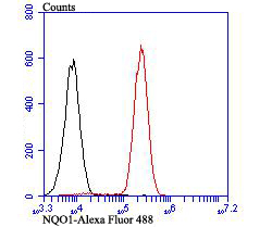
Fig4: Flow cytometric analysis of Siha cells with NQO1 antibody at 1/100 dilution (red) compared with an unlabelled control (cells without incubation with primary antibody; black). Alexa Fluor 488-conjugated goat anti-rabbit IgG was used as the secondary antibody.Application
Fig4: Flow cytometric analysis of Siha cells with NQO1 antibody at 1/100 dilution (red) compared with an unlabelled control (cells without incubation with primary antibody; black). Alexa Fluor 488-conjugated goat anti-rabbit IgG was used as the secondary antibody.Application
Fig4: Flow cytometric analysis of Siha cells with NQO1 antibody at 1/100 dilution (red) compared with an unlabelled control (cells without incubation with primary antibody; black). Alexa Fluor 488-conjugated goat anti-rabbit IgG was used as the secondary antibody.Application
Fig4: Flow cytometric analysis of Siha cells with NQO1 antibody at 1/100 dilution (red) compared with an unlabelled control (cells without incubation with primary antibody; black). Alexa Fluor 488-conjugated goat anti-rabbit IgG was used as the secondary antibody.| Product Name | NQO1 |
|---|---|
| Antibody Type | Primary Antibodies |
| Antigen Alias | Azoreductase antibody Cytochrome b 5 reductase antibody DHQU antibody DIA 4 antibody DIA4 antibody Diaphorase (NADH/NADPH) (cytochrome b 5 reductase) antibody Diaphorase (NADH/NADPH) antibody Diaphorase 4 antibody Dioxin inducible 1 antibody DT diaphorase antibody DT-diaphorase antibody DTD antibody Menadione reductase antibody NAD(P)H dehydrogenase [quinone] 1 antibody NAD(P)H dehydrogenase quinone 1 antibody NAD(P)H menadione oxidoreductase 1 dioxin inducible antibody NAD(P)H quinone dehydrogenase 1 antibody NAD(P)H: menadione oxidoreductase 1 dioxin inducible 1 antibody NAD(P)H:menadione oxidoreductase 1 antibody NAD(P)H:Quinone acceptor oxidoreductase type 1 antibody NAD(P)H:quinone oxidoreductase 1 antibody NAD(P)H:quinone oxireductase antibody NMOR 1 antibody NMOR I antibody NMOR1 antibody NMORI antibody NQO 1 antibody NQO1 antibody NQO1_HUMAN antibody Phylloquinone reductase antibody QR 1 antibody QR1 antibody Quinone reductase 1 antibody |
| Product description | NQO1, a 274 amino acid protein, is ubiquitously expressed, but the expression level varies among tissues. NQO1 gene expression is coordinately induced in response to xenobiotics, antioxidants, heavy metals and radiation. The antioxidant response element (ARE) in the NQO1 gene promoter is essential for expression and coordinated induction of NQO1. ARE activation by tert-butylhydroquinone is dependent on PI3-kinase, which lies upstream of Nrf2. Nrf2, c-Jun, Nrf1, Jun-B and Jun-D bind to the ARE and regulate expression and induction of NQO1 gene. Maf-Maf homodimers and possibly Maf-Nrf2 heterodimers play a role in negative regulation of ARE-mediated transcription, but Maf-Nrf1 heterodimers fail to bind with the NQO1 gene ARE and do not repress NQO1 transcription. |
| Immunogen | Recombinant protein within human NQO1 1-250 aa. |
| Clonality | Polyclonal |
|---|---|
| Isotype | IgG |
| Host Species | Rabbit |
| Tested Applications | WBIHCFC |
| WB:1:500-1:1,000 IHC:1:50-1:200 FC:1:50-1:100 |
|
| Species Reactivity | HumanMouseRat |
| Concentration | 1mg/ml |
| Alternative Names | Azoreductase antibody Cytochrome b 5 reductase antibody DHQU antibody DIA 4 antibody DIA4 antibody Diaphorase (NADH/NADPH) (cytochrome b 5 reductase) antibody Diaphorase (NADH/NADPH) antibody Diaphorase 4 antibody Dioxin inducible 1 antibody DT diaphorase antibody DT-diaphorase antibody DTD antibody Menadione reductase antibody NAD(P)H dehydrogenase [quinone] 1 antibody NAD(P)H dehydrogenase quinone 1 antibody NAD(P)H menadione oxidoreductase 1 dioxin inducible antibody NAD(P)H quinone dehydrogenase 1 antibody NAD(P)H: menadione oxidoreductase 1 dioxin inducible 1 antibody NAD(P)H:menadione oxidoreductase 1 antibody NAD(P)H:Quinone acceptor oxidoreductase type 1 antibody NAD(P)H:quinone oxidoreductase 1 antibody NAD(P)H:quinone oxireductase antibody NMOR 1 antibody NMOR I antibody NMOR1 antibody NMORI antibody NQO 1 antibody NQO1 antibody NQO1_HUMAN antibody Phylloquinone reductase antibody QR 1 antibody QR1 antibody Quinone reductase 1 antibody |
|---|---|
| Molecular Weight(MW) | 31 kDa |
| Cellular Localization | Cytoplasm. |
Application
Fig4: Flow cytometric analysis of Siha cells with NQO1 antibody at 1/100 dilution (red) compared with an unlabelled control (cells without incubation with primary antibody; black). Alexa Fluor 488-conjugated goat anti-rabbit IgG was used as the secondary antibody.
Application
Fig4: Flow cytometric analysis of Siha cells with NQO1 antibody at 1/100 dilution (red) compared with an unlabelled control (cells without incubation with primary antibody; black). Alexa Fluor 488-conjugated goat anti-rabbit IgG was used as the secondary antibody.
Application
Fig4: Flow cytometric analysis of Siha cells with NQO1 antibody at 1/100 dilution (red) compared with an unlabelled control (cells without incubation with primary antibody; black). Alexa Fluor 488-conjugated goat anti-rabbit IgG was used as the secondary antibody.
Application
Fig4: Flow cytometric analysis of Siha cells with NQO1 antibody at 1/100 dilution (red) compared with an unlabelled control (cells without incubation with primary antibody; black). Alexa Fluor 488-conjugated goat anti-rabbit IgG was used as the secondary antibody.| Positive Control | Siha, A549, rat kidney tissue, mouse colon tissue. |
|---|---|
| Application Notes | WB:1:500-1:1,000 IHC:1:50-1:200 FC:1:50-1:100 |
| Form | Liquid |
|---|---|
| Storage Instructions | Store at +4℃ after thawing. Aliquot store at -20℃. Avoid repeated freeze / thaw cycles. |
| Storage Buffer | 1*TBS (pH7.4), 0.5%BSA, 50%Glycerol. Preservative: 0.05% Sodium Azide. |


