Application
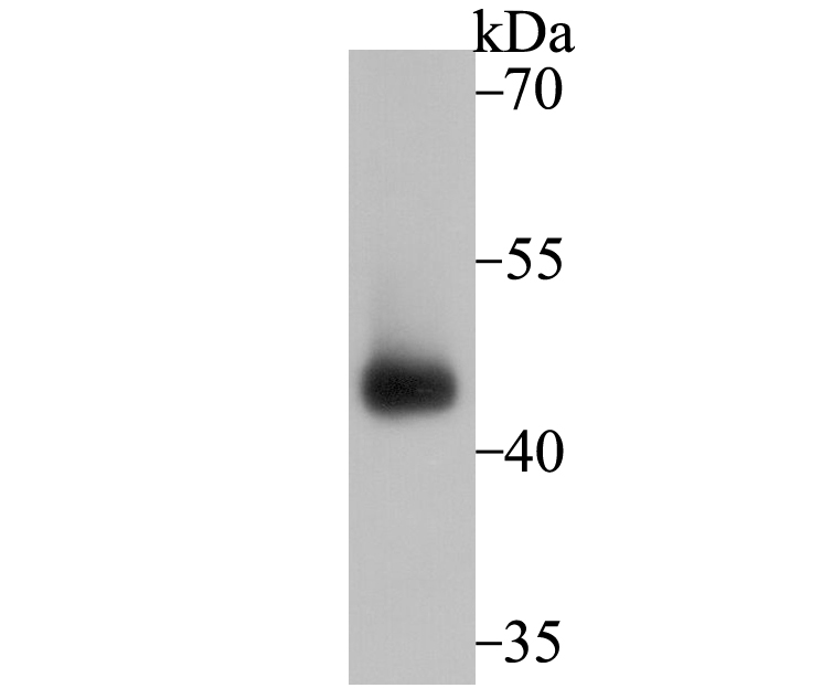
Fig1: Western blot analysis of Connexin 43 on mouse testis tissue lysate using anti-Connexin 43 antibody at 1/2,000 dilution.Application
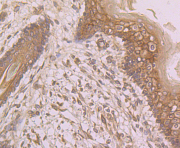
Fig2: Immunohistochemical analysis of paraffin-embedded human skin tissue using anti-Connexin 43 antibody. Counter stained with hematoxylin.Application
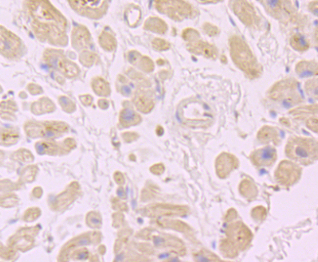
Fig3: Immunohistochemical analysis of paraffin-embedded human heart tissue using anti-Connexin 43 antibody. Counter stained with hematoxylin.Application
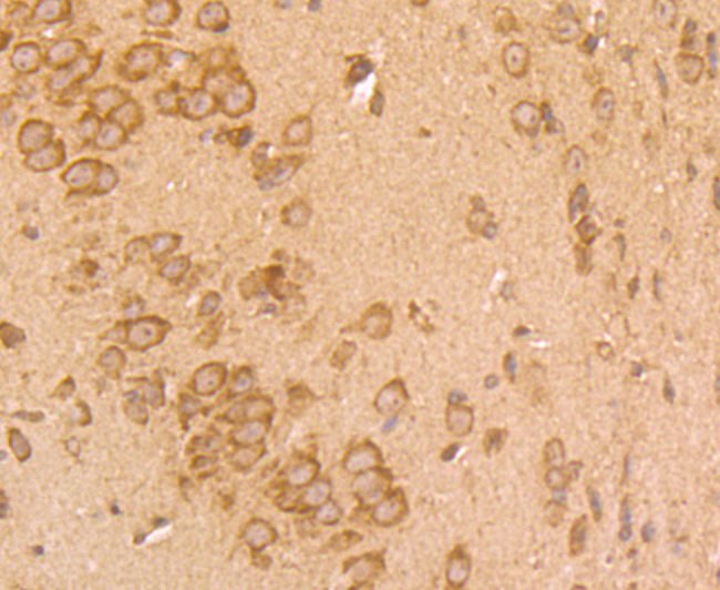
Fig4: Immunohistochemical analysis of paraffin-embedded mouse brain tissue using anti-Connexin 43 antibody. Counter stained with hematoxylin.Application
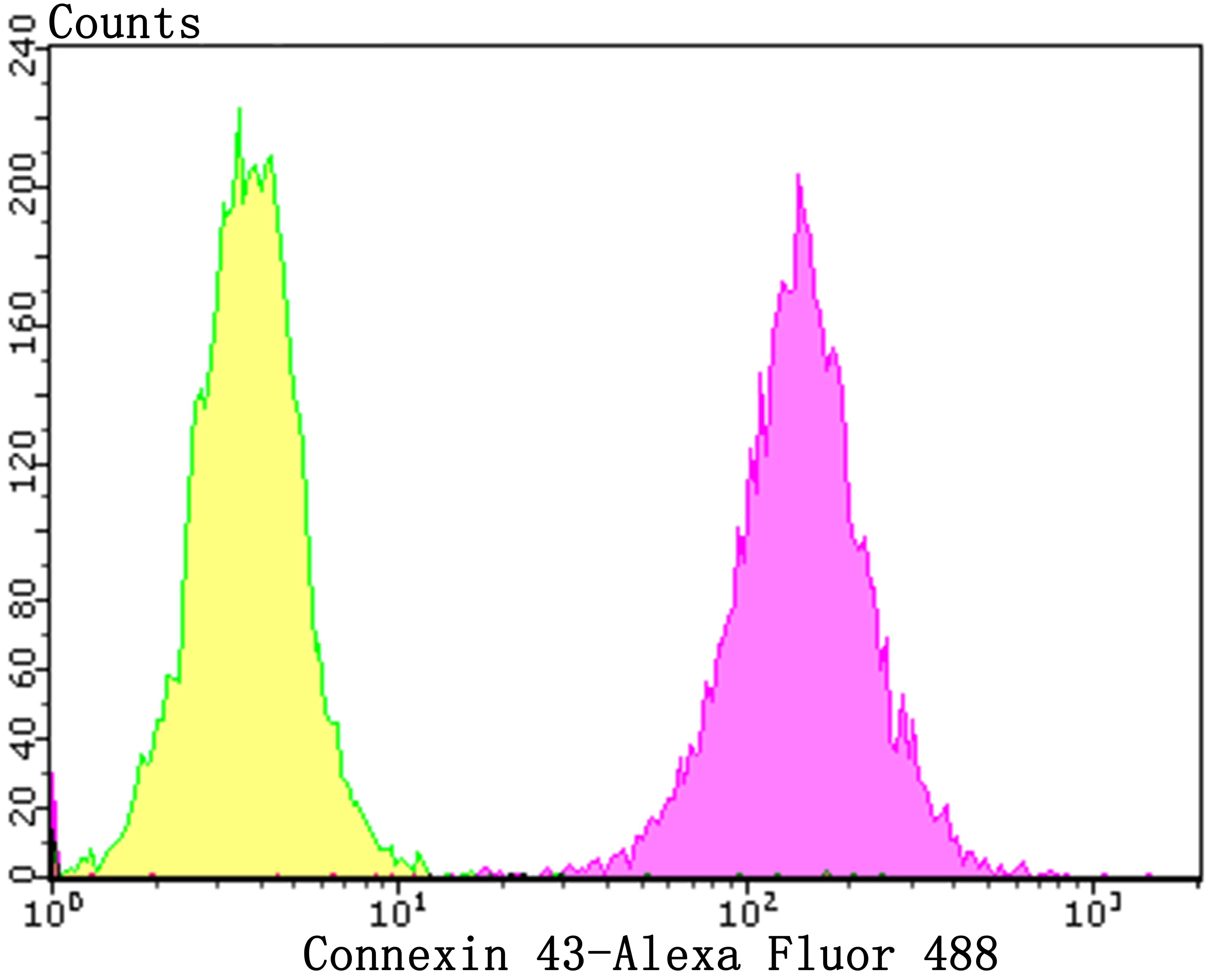
Fig5: Flow cytometric analysis of SH-SY-5Y cells with Connexin 43 antibody at 1/50 dilution (purple) compared with an unlabelled control (cells without incubation with primary antibody; yellow). Alexa Fluor 488-conjugated goat anti-rabbit IgG was used as the secondary antibody.| Product Name | Connexin 43 |
|---|---|
| Antibody Type | Primary Antibodies |
| Antigen Alias | Connexin 43 antibody Connexin-43 antibody Cx 43 antibody Cx43 antibody CXA1_HUMAN antibody DFNB38 antibody Gap junction 43 kDa heart protein antibody Gap junction alpha-1 protein antibody Gap junction protein alpha 1 43kDa (connexin 43) antibody Gap junction protein alpha 1 43kDa antibody Gap junction protein alpha like antibody GJA 1 antibody Gja1 antibody GJAL antibody ODD antibody ODDD antibody ODOD antibody SDTY3 antibody |
| Product description | The connexins are a group of gap junction proteins which form a hexamer to compose a connexon. Clusters of connexons form a gap junction through which low molecular weight proteins may diffuse from cell to cell. Several mammalian cells with malignant phenotypes exhibit decreased connexin expression and gap junction communication. In Src transformed cells, there is a decrease in gap junctional communication, which appears to be associated with tyrosine phosphorylation of connexin 43. Activated c-Src phosphorylates the C-terminal tail of connexin 43 on Tyr 265, resulting in a stable interaction between both proteins, which leads to inhibition of gap junctional communication. In addition to tyrosine phosphorylation, connexin 43 has also been shown to be phosphorylated on serine in the absence of Src kinases and on both serine and tyrosine in cells expressing Src kinases, such as c-Src and/or pp60v-Src. In human vascular endothelial cells, connexin 43 is posttranslationally modified during mitosis. Mitosis-specific phosphorylation of connexin 43 correlates with the transient loss of gap junction intercellular communication and redistribution of connexin 43. |
| Immunogen | Synthetic peptide within C-terminal human Connexin 43. |
| Clonality | Polyclonal |
|---|---|
| Isotype | IgG |
| Host Species | Rabbit |
| Tested Applications | WBIHCFC |
| WB:1:500-1:2,000 IHC:1:50-1:200 FC:1:50-1:200 |
|
| Species Reactivity | HumanMouse |
| Concentration | 1mg/ml |
| Alternative Names | Connexin 43 antibody Connexin-43 antibody Cx 43 antibody Cx43 antibody CXA1_HUMAN antibody DFNB38 antibody Gap junction 43 kDa heart protein antibody Gap junction alpha-1 protein antibody Gap junction protein alpha 1 43kDa (connexin 43) antibody Gap junction protein alpha 1 43kDa antibody Gap junction protein alpha like antibody GJA 1 antibody Gja1 antibody GJAL antibody ODD antibody ODDD antibody ODOD antibody SDTY3 antibody |
|---|---|
| Molecular Weight(MW) | 43 kDa |
| Cellular Localization | Plasma membrane. Endoplasmic reticulum. Gap junction. |
Application
Fig1: Western blot analysis of Connexin 43 on mouse testis tissue lysate using anti-Connexin 43 antibody at 1/2,000 dilution.
Application
Fig2: Immunohistochemical analysis of paraffin-embedded human skin tissue using anti-Connexin 43 antibody. Counter stained with hematoxylin.
Application
Fig3: Immunohistochemical analysis of paraffin-embedded human heart tissue using anti-Connexin 43 antibody. Counter stained with hematoxylin.
Application
Fig4: Immunohistochemical analysis of paraffin-embedded mouse brain tissue using anti-Connexin 43 antibody. Counter stained with hematoxylin.
Application
Fig5: Flow cytometric analysis of SH-SY-5Y cells with Connexin 43 antibody at 1/50 dilution (purple) compared with an unlabelled control (cells without incubation with primary antibody; yellow). Alexa Fluor 488-conjugated goat anti-rabbit IgG was used as the secondary antibody.| Positive Control | Mouse testis tissue lysate, human skin tissue, human heart tissue, mouse brain tissue, SH-SY-5Y. |
|---|---|
| Application Notes | WB:1:500-1:2,000 IHC:1:50-1:200 FC:1:50-1:200 |
| Form | Liquid |
|---|---|
| Storage Instructions | Store at +4℃ after thawing. Aliquot store at -20℃. Avoid repeated freeze / thaw cycles. |
| Storage Buffer | 1*TBS (pH7.4), 0.5%BSA, 50%Glycerol. Preservative: 0.05% Sodium Azide. |


