Application
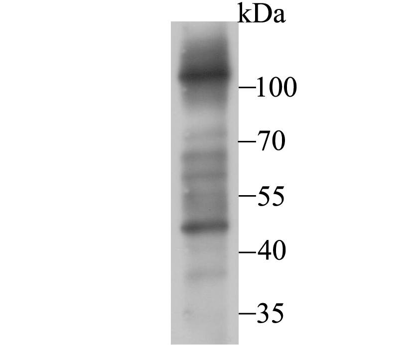
Fig1: Western blot analysis of FBXL18 on PC-3M cell lysate using anti-FBXL18 antibody at 1/500 dilution.Application
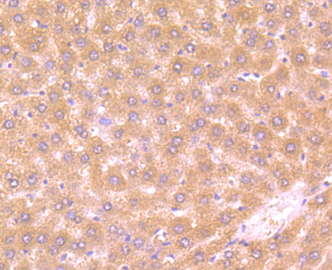
Fig2: Immunohistochemical analysis of paraffin-embedded rat liver tissue using anti-FBXL18 antibody. Counter stained with hematoxylin.Application
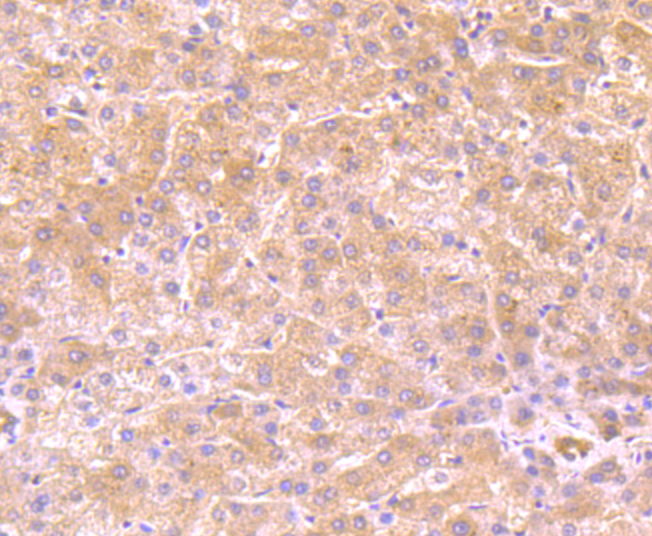
Fig3: Immunohistochemical analysis of paraffin-embedded human liver tissue using anti-FBXL18 antibody. Counter stained with hematoxylin.Application
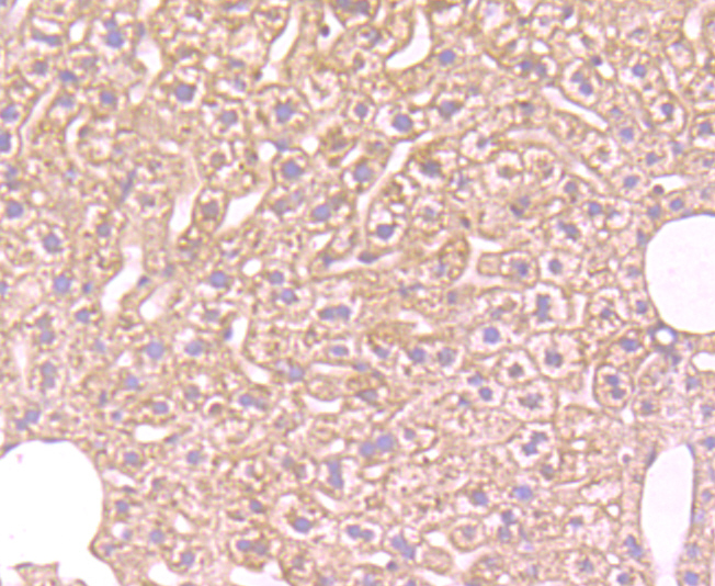
Fig4: Immunohistochemical analysis of paraffin-embedded mouse liver tissue using anti-FBXL18 antibody. Counter stained with hematoxylin.Application
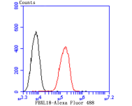
Fig5: Flow cytometric analysis of A549 cells with FBXL18 antibody at 1/100 dilution (red) compared with an unlabelled control (cells without incubation with primary antibody; black). Alexa Fluor 488-conjugated goat anti-rabbit IgG was used as the secondary antibody.| Product Name | FBXL18 |
|---|---|
| Antibody Type | Primary Antibodies |
| Antigen Alias | F box and leucine rich repeat protein 18 antibody F box/LRR repeat protein 18 antibody FBL18 antibody FBXL 18 antibody FLJ10776 antibody FLJ11467 antibody FLJ26934 antibody FLJ38075 antibody FLJ41541 antibody |
| Product description | FBL18 (F-box and leucine-rich repeat protein 18), also known as FBXL18, is an 805 amino acid protein that contains an F-box near its N-terminus, followed by several leucine-rich repeats and a transmembrane domain at the C-terminus. F-box proteins are critical components of the SCF (Skp1-CUL-1-F-box protein) type E3 ubiquitin ligase complex and are involved in substrate recognition and recruitment for ubiquitination. They are members of a larger family of proteins that are involved in the regulation of a wide variety of cellular processes (including the cell cycle, immune response, signaling cascades and developmental processes) through the targeting of proteins, such as cyclins and cyclin-dependent kinase inhibitors (CDKNs), for degradation by the proteasome after ubiquitination. FBL18 directly interacts with SKP1A p19 and CUL-1, forming a substrate-recognition component of the SCF-type E3 ubiquitin ligase complex. Four isoforms of FBL18 exist due to alternative splicing. |
| Immunogen | Synthetic peptide corresponding to human FBXL18 aa 750 to C terminus. |
| Clonality | Polyclonal |
|---|---|
| Isotype | IgG |
| Host Species | Rabbit |
| Tested Applications | WBIHCFC |
| WB:1:500 IHC:1:50-1:200 FC:1:50-1:100 |
|
| Species Reactivity | HumanMouseRat |
| Concentration | 1mg/ml |
| Alternative Names | F box and leucine rich repeat protein 18 antibody F box/LRR repeat protein 18 antibody FBL18 antibody FBXL 18 antibody FLJ10776 antibody FLJ11467 antibody FLJ26934 antibody FLJ38075 antibody FLJ41541 antibody |
|---|---|
| Molecular Weight(MW) | 88/40 kDa |
| Cellular Localization | Cytoplasm. |
| SwissProt ID | Q96ME1 |
|---|
Application
Fig1: Western blot analysis of FBXL18 on PC-3M cell lysate using anti-FBXL18 antibody at 1/500 dilution.
Application
Fig2: Immunohistochemical analysis of paraffin-embedded rat liver tissue using anti-FBXL18 antibody. Counter stained with hematoxylin.
Application
Fig3: Immunohistochemical analysis of paraffin-embedded human liver tissue using anti-FBXL18 antibody. Counter stained with hematoxylin.
Application
Fig4: Immunohistochemical analysis of paraffin-embedded mouse liver tissue using anti-FBXL18 antibody. Counter stained with hematoxylin.
Application
Fig5: Flow cytometric analysis of A549 cells with FBXL18 antibody at 1/100 dilution (red) compared with an unlabelled control (cells without incubation with primary antibody; black). Alexa Fluor 488-conjugated goat anti-rabbit IgG was used as the secondary antibody.| Positive Control | PC-3M, rat liver tissue, human liver tissue, mouse liver tissue, A549. |
|---|---|
| Application Notes | WB:1:500 IHC:1:50-1:200 FC:1:50-1:100 |
| Form | Liquid |
|---|---|
| Storage Instructions | Store at +4℃ after thawing. Aliquot store at -20℃. Avoid repeated freeze / thaw cycles. |
| Storage Buffer | 1*TBS (pH7.4), 0.5%BSA, 50%Glycerol. Preservative: 0.05% Sodium Azide. |


