Application
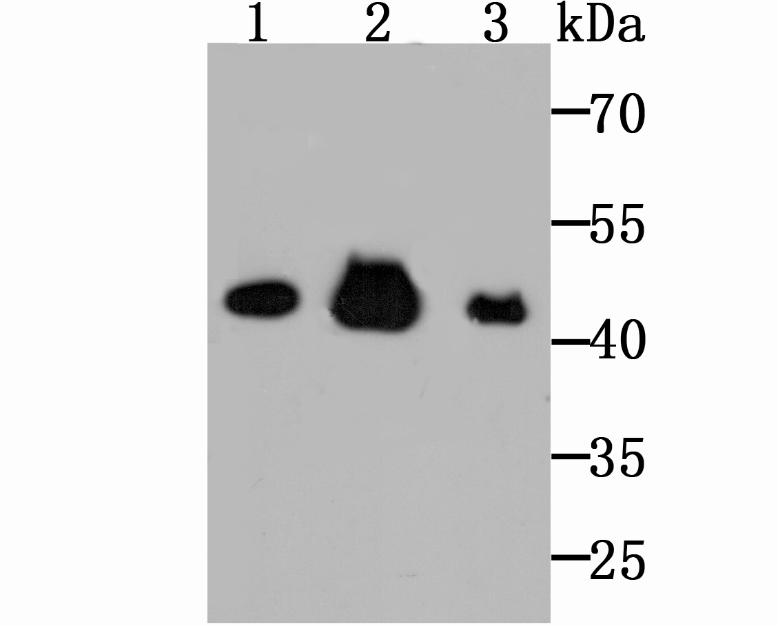
Fig1: Western blot analysis of Citrate synthetase on Hela cell using anti-Citrate synthetase antibody at 1/1,000 dilution. Positive control: Lane 1: Rat kidney tissue Lane 2: Hela Lane 3: Mouse brain tissueApplication
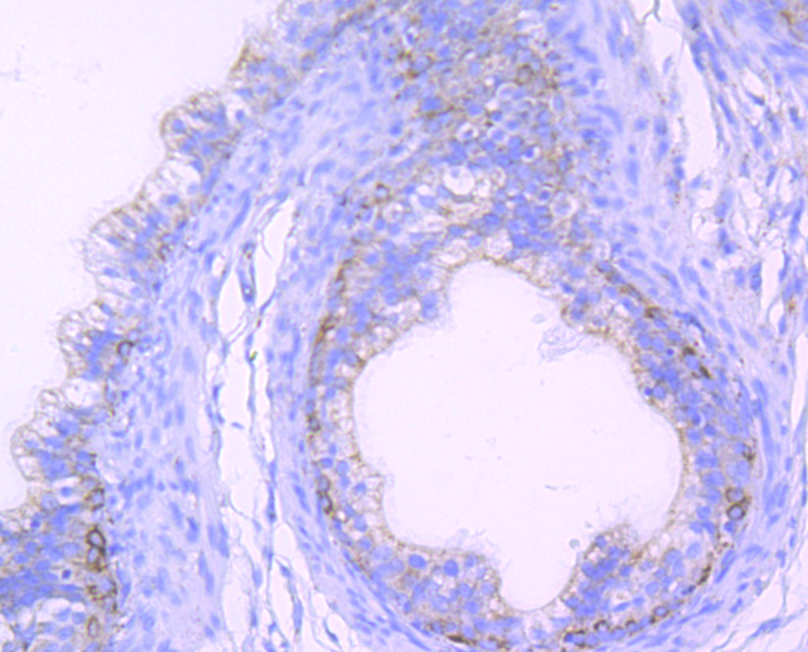
Fig2: Immunohistochemical analysis of paraffin-embedded rat epididymis tissue using anti-Citrate synthetase antibody. Counter stained with hematoxylin.Application
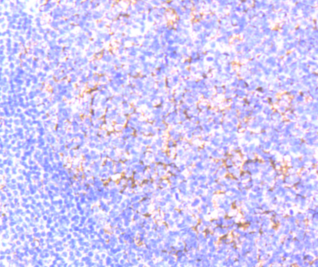
Fig3: Immunohistochemical analysis of paraffin-embedded human tonsil tissue using anti-Citrate synthetase antibody. Counter stained with hematoxylin.Application
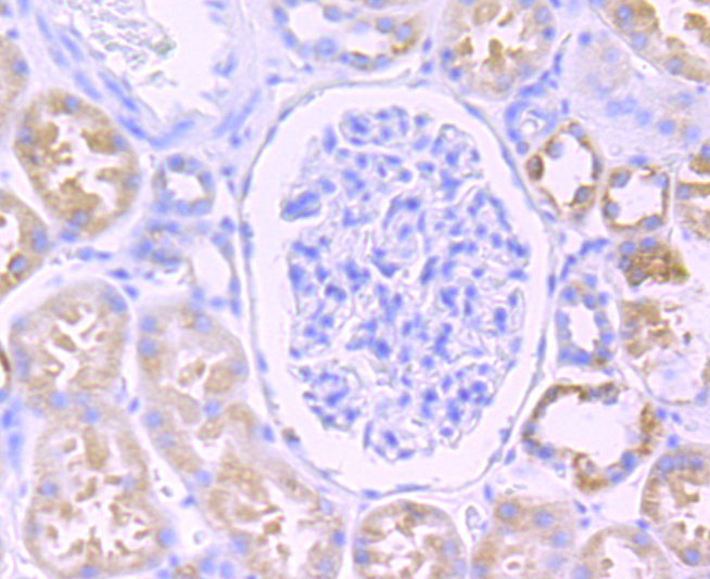
Fig4: Immunohistochemical analysis of paraffin-embedded human kidney tissue using anti-Citrate synthetase antibody. Counter stained with hematoxylin.Application
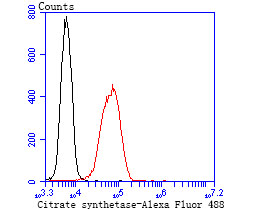
Fig5: Flow cytometric analysis of 293T cells with Citrate synthetase antibody at 1/50 dilution (blue) compared with an unlabelled control (cells without incubation with primary antibody; red). Alexa Fluor 488-conjugated goat anti rabbit IgG was used as the secondary antibody.| Product Name | Citrate synthetase [JU80-30] |
|---|---|
| Antibody Type | Primary Antibodies |
| Antigen Alias | CISY_HUMAN antibody Citrate synthase antibody Citrate synthase, mitochondrial antibody citrate synthetase antibody Cs antibody EC 2.3.3 antibody EC 2.3.3.1 antibody |
| Product description | Citrate synthase is found in nearly all cells capable of oxidative metabolism.The enzyme citrate synthase exists in nearly all living cells and stands as a pace-making enzyme in the first step of the citric acid cycle (or Krebs cycle). Citrate synthase is localized within eukaryotic cells in the mitochondrial matrix, but is encoded by nuclear DNA rather than mitochondrial. It is synthesized using cytoplasmic ribosomes, then transported into the mitochondrial matrix. Citrate synthase is commonly used as a quantitative enzyme marker for the presence of intact mitochondria. |
| Immunogen | Recombinant protein |
| Clonality | Monoclonal |
|---|---|
| Isotype | IgG |
| Host Species | Recombinant rabbit |
| Tested Applications | WBIHCFC |
| WB:1:500-1:1,000 IHC:1:50-1:100 FC:1:50-1:200 |
|
| Species Reactivity | HumanMouseRat |
| Concentration | 1mg/ml |
| Alternative Names | CISY_HUMAN antibody
Citrate synthase antibody
Citrate synthase mitochondrial antibody citrate synthetase antibody Cs antibody EC 2.3.3 antibody EC 2.3.3.1 antibody |
|---|---|
| Molecular Weight(MW) | 52 kDa |
| Cellular Localization | Mitochondrion matrix. |
Application
Fig1: Western blot analysis of Citrate synthetase on Hela cell using anti-Citrate synthetase antibody at 1/1,000 dilution. Positive control: Lane 1: Rat kidney tissue Lane 2: Hela Lane 3: Mouse brain tissue
Application
Fig2: Immunohistochemical analysis of paraffin-embedded rat epididymis tissue using anti-Citrate synthetase antibody. Counter stained with hematoxylin.
Application
Fig3: Immunohistochemical analysis of paraffin-embedded human tonsil tissue using anti-Citrate synthetase antibody. Counter stained with hematoxylin.
Application
Fig4: Immunohistochemical analysis of paraffin-embedded human kidney tissue using anti-Citrate synthetase antibody. Counter stained with hematoxylin.
Application
Fig5: Flow cytometric analysis of 293T cells with Citrate synthetase antibody at 1/50 dilution (blue) compared with an unlabelled control (cells without incubation with primary antibody; red). Alexa Fluor 488-conjugated goat anti rabbit IgG was used as the secondary antibody.| Positive Control | Mouse brain tissue lysate, rat kidney tissue lysate, Hela, rat epididymis tissue, human tonsil tissue, human kidney tissue, 293T. |
|---|---|
| Application Notes | WB:1:500-1:1,000 IHC:1:50-1:100 FC:1:50-1:200 |
| Form | Liquid |
|---|---|
| Storage Instructions | Store at +4℃ after thawing. Aliquot store at -20℃ or -80℃. Avoid repeated freeze / thaw cycles. |
| Storage Buffer | 1*TBS (pH7.4), 1%BSA, 40%Glycerol. Preservative: 0.05% Sodium Azide. |


