Application
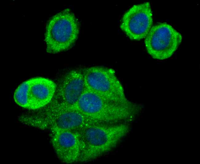
Fig1: ICC staining TAK1 in MCF-7 cells (green). The nuclear counter stain is DAPI (blue). Cells were fixed in paraformaldehyde, permeabilised with 0.25% Triton X100/PBS.Application
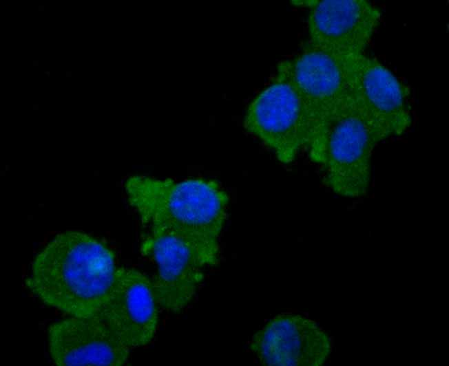
Fig2: ICC staining TAK1 in N2A cells (green). The nuclear counter stain is DAPI (blue). Cells were fixed in paraformaldehyde, permeabilised with 0.25% Triton X100/PBS.Application
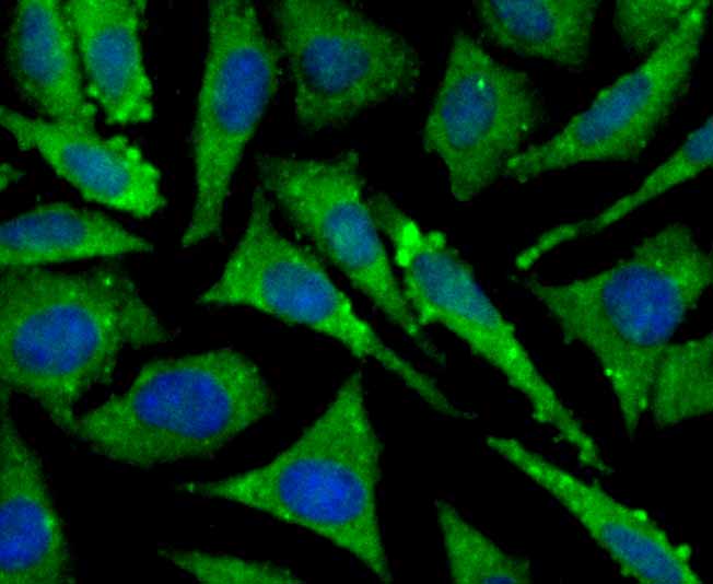
Fig3: ICC staining TAK1 in SH-SY-5Y cells (green). The nuclear counter stain is DAPI (blue). Cells were fixed in paraformaldehyde, permeabilised with 0.25% Triton X100/PBS.Application
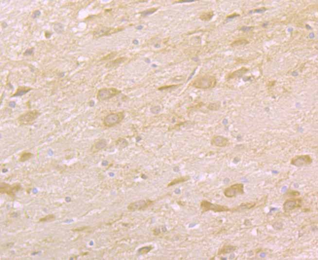
Fig4: Immunohistochemical analysis of paraffin-embedded rat brain tissue using anti-TAK1 antibody. Counter stained with hematoxylin.Application
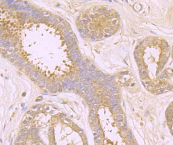
Fig5: Immunohistochemical analysis of paraffin-embedded human breast tissue using anti-TAK1 antibody. Counter stained with hematoxylin.Application
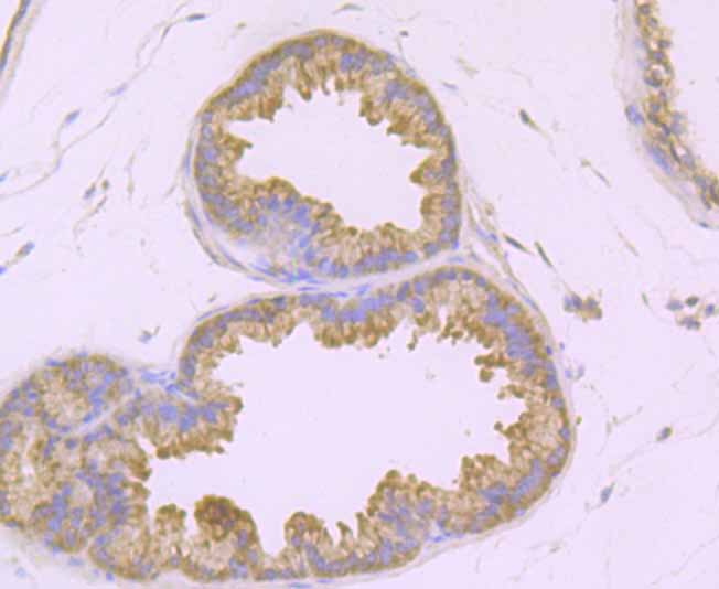
Fig6: Immunohistochemical analysis of paraffin-embedded mouse prostate tissue using anti-TAK1 antibody. Counter stained with hematoxylin.Application
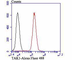
Fig7: Flow cytometric analysis of K562 cells with TAK1 antibody at 1/100 dilution (red) compared with an unlabelled control (cells without incubation with primary antibody; black). Alexa Fluor 488-conjugated goat anti-rabbit IgG was used as the secondary antibody.| Product Name | TAK1 |
|---|---|
| Antibody Type | Primary Antibodies |
| Antigen Alias | M3K7_HUMAN antibody MAP3K 7 antibody Map3k7 antibody MEKK7 antibody Mitogen activated protein kinase kinase kinase 7 antibody Mitogen-activated protein kinase kinase kinase 7 antibody TAK1 antibody TGF beta activated kinase 1 antibody TGF-beta-activated kinase 1 antibody TGF1a antibody Transforming growth factor beta activated kinase 1 antibody Transforming growth factor-beta-activated kinase 1 antibody |
| Product description | nases, ribosomal S6 kinase (Rsk) and Raf-1. Raf-1 is a protein with intrinsic kinase activity towards serine/ threonine residues and that is widely expressed in many tissue types and cell lines. Raf-1 activation is dependent on the small molecular weight GTPase Ras, but the means by which this activation occurs is poorly understood. Two proteins putatively involved in this process are Ksr-1 and Tak1. Ksr-1 (kinase sup-pressor of Ras) is a novel Raf-related protein kinase whose function is required for Ras signal transduction. Whether Ksr-1 lies directly downstream of Ras or acts in a parallel pathway is not yet known. Tak1 (TGFβ-activated kinase) has been shown to participate in the activation of the MAP kinase family in response to TGFβ stimulation. |
| Immunogen | Synthetic peptide within Human TAK1 aa 560 to the C-terminus . |
| Clonality | Polyclonal |
|---|---|
| Isotype | IgG |
| Host Species | Rabbit |
| Tested Applications | ICCIHCFC |
| ICC:1:50-1:200 IHC:1:50-1:200 FC:1:50-1:100 |
|
| Species Reactivity | HumanMouseRat |
| Concentration | 1mg/ml |
| Alternative Names | M3K7_HUMAN antibody MAP3K 7 antibody Map3k7 antibody MEKK7 antibody Mitogen activated protein kinase kinase kinase 7 antibody Mitogen-activated protein kinase kinase kinase 7 antibody TAK1 antibody TGF beta activated kinase 1 antibody TGF-beta-activated kinase 1 antibody TGF1a antibody Transforming growth factor beta activated kinase 1 antibody Transforming growth factor-beta-activated kinase 1 antibody |
|---|---|
| Molecular Weight(MW) | 67 kDa |
| Cellular Localization | Cytoplasm. Membrane. |
Application
Fig1: ICC staining TAK1 in MCF-7 cells (green). The nuclear counter stain is DAPI (blue). Cells were fixed in paraformaldehyde, permeabilised with 0.25% Triton X100/PBS.
Application
Fig2: ICC staining TAK1 in N2A cells (green). The nuclear counter stain is DAPI (blue). Cells were fixed in paraformaldehyde, permeabilised with 0.25% Triton X100/PBS.
Application
Fig3: ICC staining TAK1 in SH-SY-5Y cells (green). The nuclear counter stain is DAPI (blue). Cells were fixed in paraformaldehyde, permeabilised with 0.25% Triton X100/PBS.
Application
Fig4: Immunohistochemical analysis of paraffin-embedded rat brain tissue using anti-TAK1 antibody. Counter stained with hematoxylin.
Application
Fig5: Immunohistochemical analysis of paraffin-embedded human breast tissue using anti-TAK1 antibody. Counter stained with hematoxylin.
Application
Fig6: Immunohistochemical analysis of paraffin-embedded mouse prostate tissue using anti-TAK1 antibody. Counter stained with hematoxylin.
Application
Fig7: Flow cytometric analysis of K562 cells with TAK1 antibody at 1/100 dilution (red) compared with an unlabelled control (cells without incubation with primary antibody; black). Alexa Fluor 488-conjugated goat anti-rabbit IgG was used as the secondary antibody.| Positive Control | MCF-7, N2A, SH-SY-5Y, rat brain tissue, human breast tissue, mouse prostate tissue, K562. |
|---|---|
| Application Notes | ICC:1:50-1:200 IHC:1:50-1:200 FC:1:50-1:100 |
| Form | Liquid |
|---|---|
| Storage Instructions | Store at +4℃ after thawing. Aliquot store at -20℃. Avoid repeated freeze / thaw cycles. |
| Storage Buffer | 1*TBS (pH7.4), 0.5%BSA, 50%Glycerol. Preservative: 0.05% Sodium Azide. |


