Application
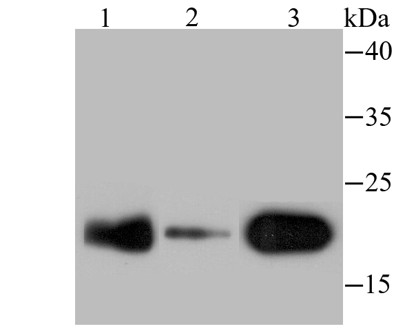
Fig1: Western blot analysis of p23 on different lysates using anti-p23 antibody at 1/500 dilution. Positive control: Lane 1: Mouse brain tissue Lane 2: SK-Br-3 Lane 3: Rat lung tissueApplication
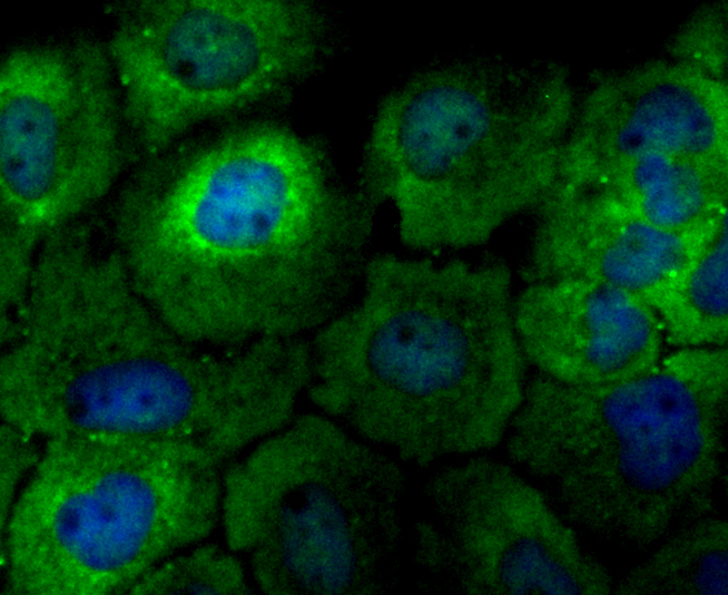
Fig2: ICC staining p23 in A431 cells (green). The nuclear counter stain is DAPI (blue). Cells were fixed in paraformaldehyde, permeabilised with 0.25% Triton X100/PBS.Application
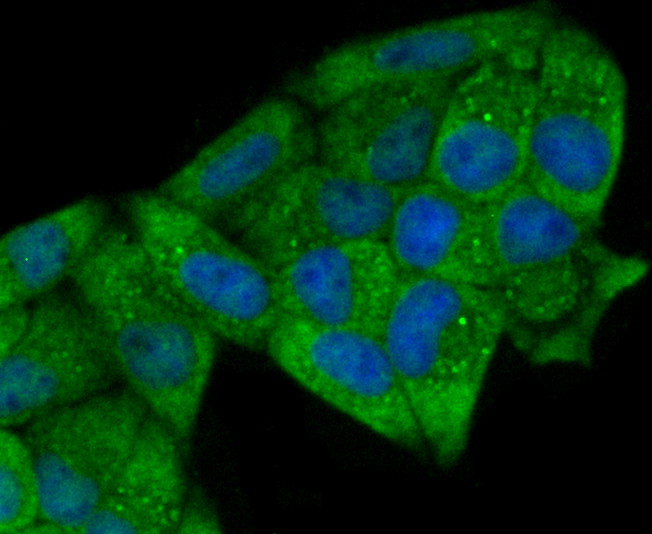
Fig3: ICC staining p23 in Hela cells (green). The nuclear counter stain is DAPI (blue). Cells were fixed in paraformaldehyde, permeabilised with 0.25% Triton X100/PBS.Application
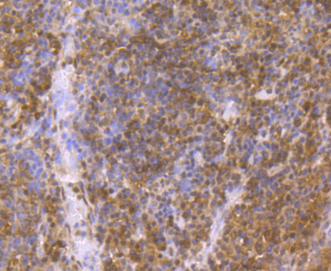
Fig4: Immunohistochemical analysis of paraffin-embedded human tonsil tissue using anti-p23 antibody. Counter stained with hematoxylin.Application
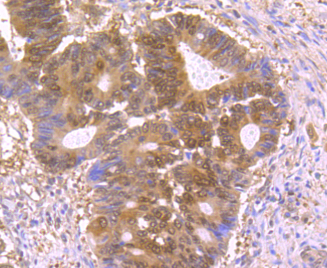
Fig5: Immunohistochemical analysis of paraffin-embedded human colon cancer tissue using anti-p23 antibody. Counter stained with hematoxylin.Application
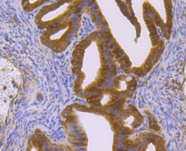
Fig6: Immunohistochemical analysis of paraffin-embedded mouse fallopian tube tissue using anti-p23 antibody. Counter stained with hematoxylin.Application
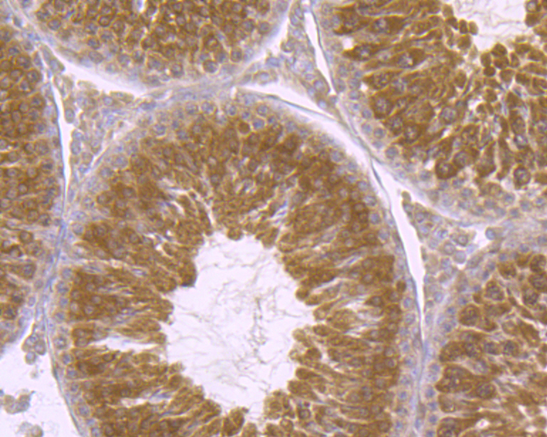
Fig7: Immunohistochemical analysis of paraffin-embedded rat testis tissue using anti-p23 antibody. Counter stained with hematoxylin.| Product Name | p23 [JU09-31] |
|---|---|
| Antibody Type | Primary Antibodies |
| Antigen Alias | Co chaperone p23 antibody cPGES antibody Cytosolic prostaglandin E synthase antibody Cytosolic prostaglandin E2 synthase antibody Hsp90 co chaperone antibody Hsp90 co-chaperone antibody P23 antibody Progesterone receptor complex antibody Progesterone receptor complex p23 antibody Prostaglandin E synthase 3 (cytosolic) antibody Prostaglandin E synthase 3 antibody PTGES 3 antibody PTGES3 antibody Sid 3177 antibody TEBP antibody TEBP_HUMAN antibody Telomerase binding protein p23 antibody Telomerase-binding protein p23 antibody Unactive progesterone receptor 23 kD antibody |
| Product description | P23, also known as PTGES3 (prostaglandin E synthase 3) or TEBP (telomerase-binding protein p23), is a ubiquitously expressed protein that functions as a cochaperone and plays an important role in signal transduction. One of several proteins in the HSP 90-based molecular chaperone complex, P23 promotes the breakdown of transcriptional regulatory complexes by disrupting receptor-mediated transcriptional activation. P23 acts in a hormone-dependent manner to chaperone estrogen receptor alpha (ERα), a steroid complex, to its mature form and to regulate the expression of ERα-related genes. Localized to the cytoplasm, P23 interacts with the glucocorticoid receptor (GR) and, through disassembly of the GR transcription machinery, is thought to inhibit GR-dependent transcription. The involvement of P23 in various steroid receptor-mediated pathways suggests close involvement in signal transduction and regulation of cellular processes. Upregulation of P23 is implicated in the invasion and metastasis of various cancers. |
| Immunogen | Recombinant protein |
| Clonality | Monoclonal |
|---|---|
| Isotype | IgG |
| Host Species | Recombinant rabbit |
| Tested Applications | WBICCIHCIP |
| WB:1:500-1:2,000 ICC:1:50-1:200 IHC:1:100-1:500 IP:1:10-1:50 |
|
| Species Reactivity | HumanMouseRat |
| Concentration | 1mg/ml |
| Alternative Names | Co chaperone p23 antibody cPGES antibody Cytosolic prostaglandin E synthase antibody Cytosolic prostaglandin E2 synthase antibody Hsp90 co chaperone antibody Hsp90 co-chaperone antibody P23 antibody Progesterone receptor complex antibody Progesterone receptor complex p23 antibody Prostaglandin E synthase 3 (cytosolic) antibody Prostaglandin E synthase 3 antibody PTGES 3 antibody PTGES3 antibody Sid 3177 antibody TEBP antibody TEBP_HUMAN antibody Telomerase binding protein p23 antibody Telomerase-binding protein p23 antibody Unactive progesterone receptor 23 kD antibody |
|---|---|
| Molecular Weight(MW) | 23 kDa |
| Cellular Localization | Cytoplasm. |
Application
Fig1: Western blot analysis of p23 on different lysates using anti-p23 antibody at 1/500 dilution. Positive control: Lane 1: Mouse brain tissue Lane 2: SK-Br-3 Lane 3: Rat lung tissue
Application
Fig2: ICC staining p23 in A431 cells (green). The nuclear counter stain is DAPI (blue). Cells were fixed in paraformaldehyde, permeabilised with 0.25% Triton X100/PBS.
Application
Fig3: ICC staining p23 in Hela cells (green). The nuclear counter stain is DAPI (blue). Cells were fixed in paraformaldehyde, permeabilised with 0.25% Triton X100/PBS.
Application
Fig4: Immunohistochemical analysis of paraffin-embedded human tonsil tissue using anti-p23 antibody. Counter stained with hematoxylin.
Application
Fig5: Immunohistochemical analysis of paraffin-embedded human colon cancer tissue using anti-p23 antibody. Counter stained with hematoxylin.
Application
Fig6: Immunohistochemical analysis of paraffin-embedded mouse fallopian tube tissue using anti-p23 antibody. Counter stained with hematoxylin.
Application
Fig7: Immunohistochemical analysis of paraffin-embedded rat testis tissue using anti-p23 antibody. Counter stained with hematoxylin.| Positive Control | Mouse brain tissue lysate, SK-Br-3, rat lung tissue lysate, A431, Hela, human tonsil tissue, human colon cancer tissue, mouse fallopian tube tissue, rat testis tissue. |
|---|---|
| Application Notes | WB:1:500-1:2,000 ICC:1:50-1:200 IHC:1:100-1:500 IP:1:10-1:50 |
| Form | Liquid |
|---|---|
| Storage Instructions | Store at +4℃ after thawing. Aliquot store at -20℃ or -80℃. Avoid repeated freeze / thaw cycles. |
| Storage Buffer | 1*TBS (pH7.4), 1%BSA, 40%Glycerol. Preservative: 0.05% Sodium Azide. |


