Application
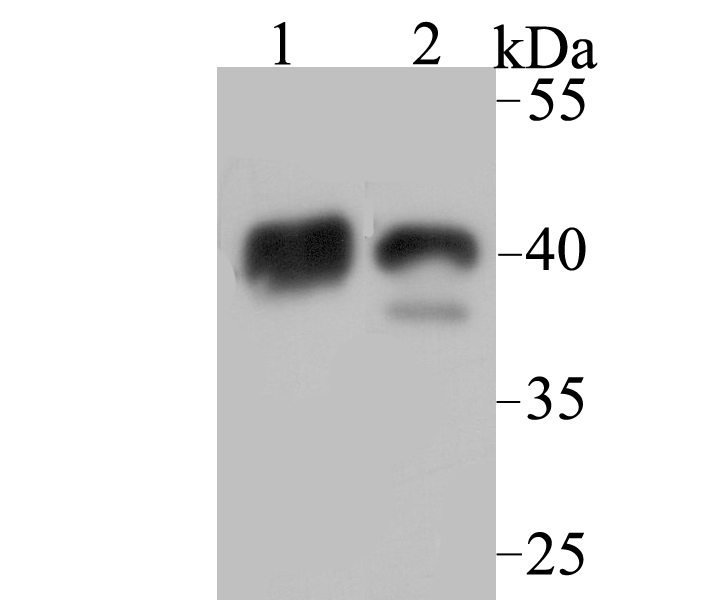
Fig1: Western blot analysis of NDRG2 on mouse brain (1) and mouse skeletal muscle (2) tissue lysate using anti-NDRG2 antibody at 1/1,000 dilution.Application
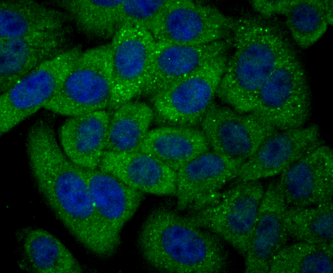
Fig2: ICC staining NDRG2 in HepG2 cells (green). The nuclear counter stain is DAPI (blue). Cells were fixed in paraformaldehyde, permeabilised with 0.25% Triton X100/PBS.Application
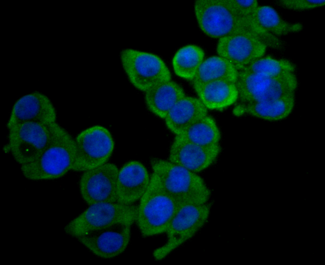
Fig3: ICC staining NDRG2 in LOVO cells (green). The nuclear counter stain is DAPI (blue). Cells were fixed in paraformaldehyde, permeabilised with 0.25% Triton X100/PBS.Application
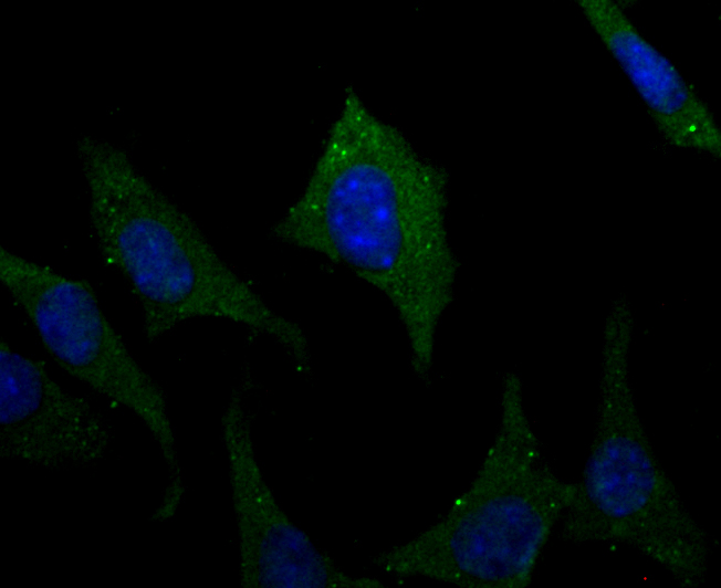
Fig4: ICC staining NDRG2 in SH-SY-5Y cells (green). The nuclear counter stain is DAPI (blue). Cells were fixed in paraformaldehyde, permeabilised with 0.25% Triton X100/PBS.Application
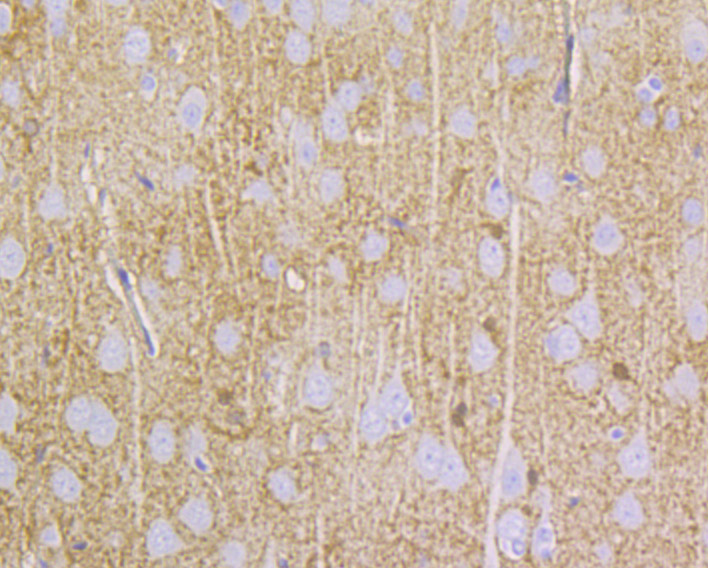
Fig5: Immunohistochemical analysis of paraffin-embedded rat brain tissue using anti-NDRG2 antibody. Counter stained with hematoxylin.Application
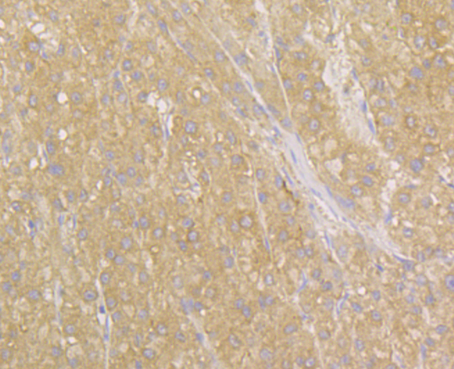
Fig6: Immunohistochemical analysis of paraffin-embedded human liver tissue using anti-NDRG2 antibody. Counter stained with hematoxylin.Application
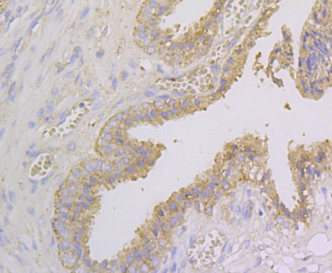
Fig7: Immunohistochemical analysis of paraffin-embedded human prostate tissue using anti-NDRG2 antibody. Counter stained with hematoxylin.Application
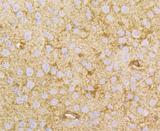
Fig8: Immunohistochemical analysis of paraffin-embedded mouse brain tissue using anti-NDRG2 antibody. Counter stained with hematoxylin.Application
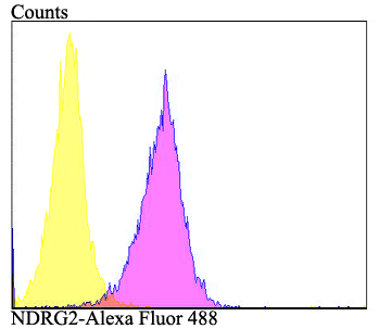
Fig9: Flow cytometric analysis of HepG2 cells with NDRG2 antibody at 1/100 dilution (purple) compared with an unlabelled control (cells without incubation with primary antibody; yellow). Alexa Fluor 488-conjugated goat anti-rabbit IgG was used as the secondary antibody.| Product Name | NDRG2 |
|---|---|
| Antibody Type | Primary Antibodies |
| Antigen Alias | Antidepressant related protein ADRG123 antibody Cytoplasmic protein Ndr1 antibody DKFZp781G1938 antibody FLJ25522 antibody KIAA1248 antibody N myc downstream regulated gene 2 antibody N myc downstream regulator 2 antibody NDR1 related protein NDR2 antibody NDRG 2 antibody NDRG family member 2 antibody NDRG1 related protein antibody NDRG2 antibody NDRG2_HUMAN antibody Protein NDRG2 antibody Protein Syld709613 antibody SYLD antibody Syld709613 protein antibody |
| Product description | The N-myc downstream regulated gene (NDRG) family is comprised of four members, namely NDRG1, NDRG2, NDRG3 and NDRG4, all of which share 57-65% homology. NDRG2 (NDRG family member 2), also known as SYLD, is a 371 amino acid protein that localizes to both the cytoplasm and the perinuclear region in neurons. Expressed at high levels in heart, brain, dendritic cells, salivary gland and skeletal muscle and at lower levels in liver and kidney, NDRG2 is thought to be involved in dendritic and neuronal cell differentiation and outgrowth. Additionally, NDRG2 expression is downregulated in a variety of carcinomas, including liver cancer, pancreatic cancer and meningioma, suggesting a possible role for NDRG2 in tumor suppression. NDRG2 is found in brain lesions of Alzheimer Disease (AD)-affected patients and is thought to be associated with the progression of AD. Five isoforms of NDRG2 exist due to alternative splicing events. |
| Immunogen | Recombinant protein within human NDRG2 1-150 aa. |
| Clonality | Polyclonal |
|---|---|
| Isotype | IgG |
| Host Species | Rabbit |
| Tested Applications | WBICCIHCFC |
| WB:1:500-1:1,000 ICC:1:50-1:200 IHC:1:50-1:100 FC:1:50-1:100 |
|
| Species Reactivity | HumanMouseRat |
| Concentration | 1mg/ml |
| Alternative Names | Antidepressant related protein ADRG123 antibody Cytoplasmic protein Ndr1 antibody DKFZp781G1938 antibody FLJ25522 antibody KIAA1248 antibody N myc downstream regulated gene 2 antibody N myc downstream regulator 2 antibody NDR1 related protein NDR2 antibody NDRG 2 antibody NDRG family member 2 antibody NDRG1 related protein antibody NDRG2 antibody NDRG2_HUMAN antibody Protein NDRG2 antibody Protein Syld709613 antibody SYLD antibody Syld709613 protein antibody |
|---|---|
| Molecular Weight(MW) | 41 kDa |
| Cellular Localization | Cytoplasm. |
Application
Fig1: Western blot analysis of NDRG2 on mouse brain (1) and mouse skeletal muscle (2) tissue lysate using anti-NDRG2 antibody at 1/1,000 dilution.
Application
Fig2: ICC staining NDRG2 in HepG2 cells (green). The nuclear counter stain is DAPI (blue). Cells were fixed in paraformaldehyde, permeabilised with 0.25% Triton X100/PBS.
Application
Fig3: ICC staining NDRG2 in LOVO cells (green). The nuclear counter stain is DAPI (blue). Cells were fixed in paraformaldehyde, permeabilised with 0.25% Triton X100/PBS.
Application
Fig4: ICC staining NDRG2 in SH-SY-5Y cells (green). The nuclear counter stain is DAPI (blue). Cells were fixed in paraformaldehyde, permeabilised with 0.25% Triton X100/PBS.
Application
Fig5: Immunohistochemical analysis of paraffin-embedded rat brain tissue using anti-NDRG2 antibody. Counter stained with hematoxylin.
Application
Fig6: Immunohistochemical analysis of paraffin-embedded human liver tissue using anti-NDRG2 antibody. Counter stained with hematoxylin.
Application
Fig7: Immunohistochemical analysis of paraffin-embedded human prostate tissue using anti-NDRG2 antibody. Counter stained with hematoxylin.
Application
Fig8: Immunohistochemical analysis of paraffin-embedded mouse brain tissue using anti-NDRG2 antibody. Counter stained with hematoxylin.
Application
Fig9: Flow cytometric analysis of HepG2 cells with NDRG2 antibody at 1/100 dilution (purple) compared with an unlabelled control (cells without incubation with primary antibody; yellow). Alexa Fluor 488-conjugated goat anti-rabbit IgG was used as the secondary antibody.| Positive Control | Mouse brain tissue, mouse skeletal muscle tissue, HepG2, LOVO, SH-SY-5Y, rat brain tissue, human liver tissue, human prostate tissue. |
|---|---|
| Application Notes | WB:1:500-1:1,000 ICC:1:50-1:200 IHC:1:50-1:100 FC:1:50-1:100 |
| Form | Liquid |
|---|---|
| Storage Instructions | Store at +4℃ after thawing. Aliquot store at -20℃. Avoid repeated freeze / thaw cycles. |
| Storage Buffer | 1*TBS (pH7.4), 0.5%BSA, 50%Glycerol. Preservative: 0.05% Sodium Azide. |


