Application
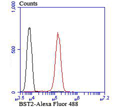
Fig4: Flow cytometric analysis of Jurkat cells with BST2 antibody at 1/100 dilution (red) compared with an unlabelled control (cells without incubation with primary antibody; black).Application
Fig4: Flow cytometric analysis of Jurkat cells with BST2 antibody at 1/100 dilution (red) compared with an unlabelled control (cells without incubation with primary antibody; black).Application
Fig4: Flow cytometric analysis of Jurkat cells with BST2 antibody at 1/100 dilution (red) compared with an unlabelled control (cells without incubation with primary antibody; black).Application
Fig4: Flow cytometric analysis of Jurkat cells with BST2 antibody at 1/100 dilution (red) compared with an unlabelled control (cells without incubation with primary antibody; black).| Product Name | BST2 [JU11-93] |
|---|---|
| Antibody Type | Primary Antibodies |
| Antigen Alias | Bone marrow stromal antigen 2 antibody Bone marrow stromal cell antigen 2 antibody Bone marrow stromal cell antigen antibody BST 2 antibody BST-2 antibody BST2 antibody BST2_HUMAN antibody CD 317 antibody CD317 antibody CD317 antigen antibody HM1.24 antigen antibody NPC A 7 antibody Tetherin antibody |
| Product description | May be involved in the sorting of secreted proteins. May be involved in pre-B-cell growth. Antiretroviral defense protein, that blocks release of retrovirus from the cell surface. Depleted unpon HIV-1 infection by viral VPU protein through 20S proteasome degradation. Depleted upon infection by human Kaposi's sarcoma-associated herpesvirus (KSHV) through ubiquitination and subsequent degradation. May play a role in B-cell activation in rheumatoid arthritis. |
| Immunogen | Recombinant protein |
| Clonality | Monoclonal |
|---|---|
| Isotype | IgG |
| Host Species | Recombinant rabbit |
| Tested Applications | WBIHCFC |
| WB:1:500 IHC:1:50-1:200 FC:1:50-1:100 |
|
| Species Reactivity | Human |
| Concentration | 1mg/ml |
| Alternative Names | Bone marrow stromal antigen 2 antibody Bone marrow stromal cell antigen 2 antibody Bone marrow stromal cell antigen antibody BST 2 antibody BST-2 antibody BST2 antibody BST2_HUMAN antibody CD 317 antibody CD317 antibody CD317 antigen antibody HM1.24 antigen antibody NPC A 7 antibody Tetherin antibody |
|---|---|
| Molecular Weight(MW) | 40 kDa |
| Cellular Localization | Golgi apparatus. Cell membrane. |
| SwissProt ID | Q10589 |
|---|
Application
Fig4: Flow cytometric analysis of Jurkat cells with BST2 antibody at 1/100 dilution (red) compared with an unlabelled control (cells without incubation with primary antibody; black).
Application
Fig4: Flow cytometric analysis of Jurkat cells with BST2 antibody at 1/100 dilution (red) compared with an unlabelled control (cells without incubation with primary antibody; black).
Application
Fig4: Flow cytometric analysis of Jurkat cells with BST2 antibody at 1/100 dilution (red) compared with an unlabelled control (cells without incubation with primary antibody; black).
Application
Fig4: Flow cytometric analysis of Jurkat cells with BST2 antibody at 1/100 dilution (red) compared with an unlabelled control (cells without incubation with primary antibody; black).| Positive Control | Hela, SiHa, human colon cancer tissue, human kidney tissue, Jurkat. |
|---|---|
| Application Notes | WB:1:500 IHC:1:50-1:200 FC:1:50-1:100 |
| Form | Liquid |
|---|---|
| Storage Instructions | Store at +4℃ after thawing. Aliquot store at -20℃ or -80℃. Avoid repeated freeze / thaw cycles. |
| Storage Buffer | 1*TBS (pH7.4), 1%BSA, 40%Glycerol. Preservative: 0.05% Sodium Azide. |


