
Application
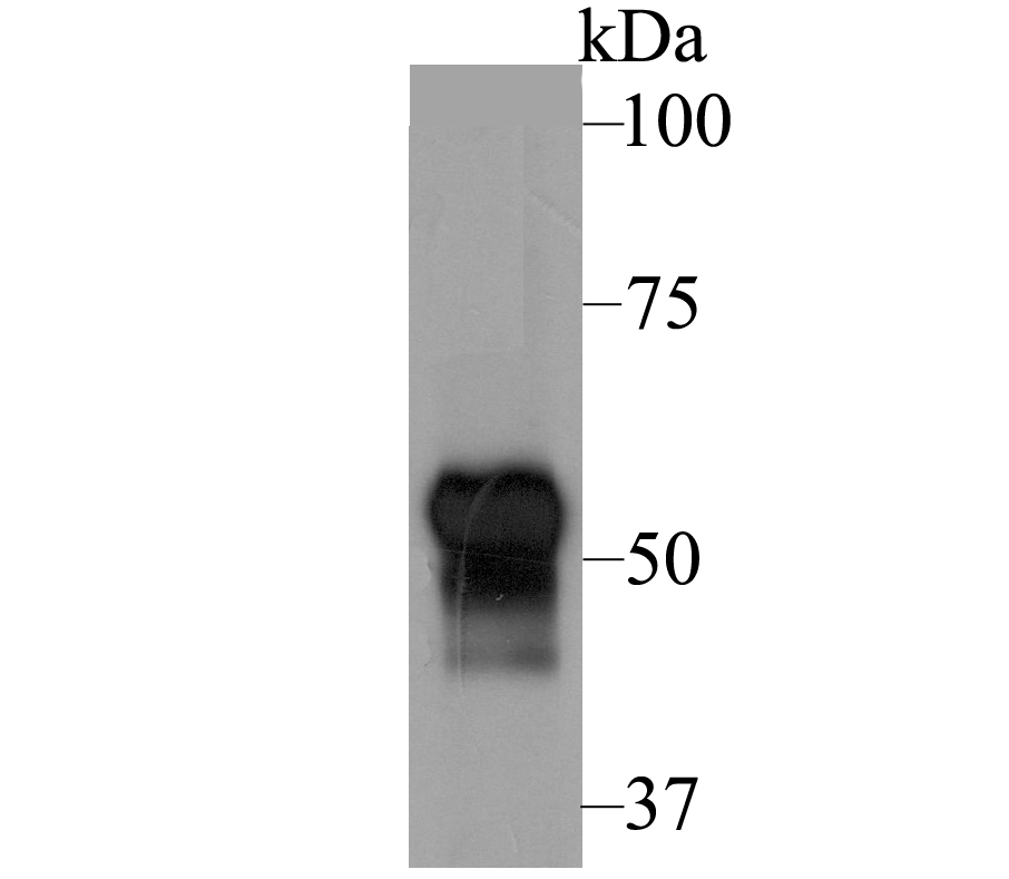
Fig1: Western blot analysis of Fibulin 5 on human skin tissue lysate using anti-Fibulin 5 antibody at 1/1,000 dilution.Application
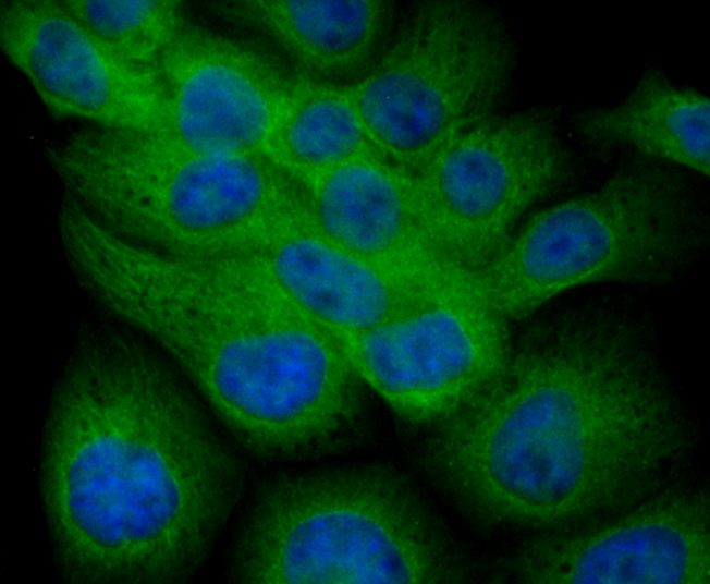
Fig2: ICC staining Fibulin 5 in JAR cells (green). The nuclear counter stain is DAPI (blue). Cells were fixed in paraformaldehyde, permeabilised with 0.25% Triton X100/PBS.Application
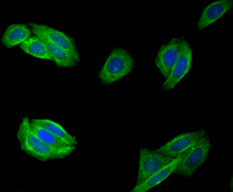
Fig3: ICC staining Fibulin 5 in SiHa cells (green). The nuclear counter stain is DAPI (blue). Cells were fixed in paraformaldehyde, permeabilised with 0.25% Triton X100/PBS.Application
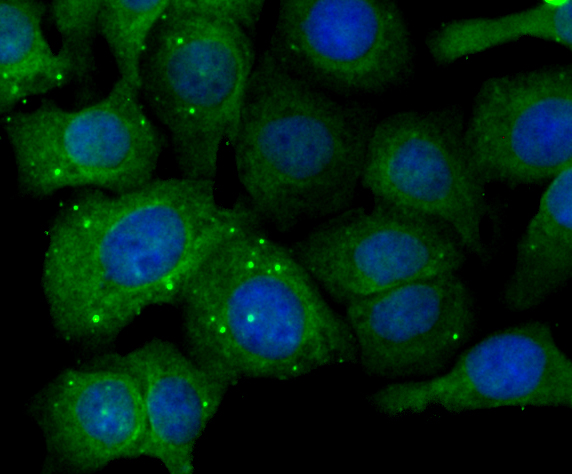
Fig4: ICC staining Fibulin 5 in SKOV-3 cells (green). The nuclear counter stain is DAPI (blue). Cells were fixed in paraformaldehyde, permeabilised with 0.25% Triton X100/PBS.Application
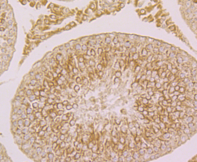
Fig5: Immunohistochemical analysis of paraffin-embedded rat testis tissue using anti-Fibulin 5 antibody. Counter stained with hematoxylin.Application
Fig6: Immunohistochemical analysis of paraffin-embedded mouse ovary tissue using anti-Fibulin 5 antibody. Counter stained with hematoxylin.Application
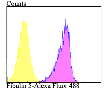
Fig7: Flow cytometric analysis of SiHa cells with Fibulin 5 antibody at 1/100 dilution (purple) compared with an unlabelled control (cells without incubation with primary antibody; yellow). Alexa Fluor 488-conjugated goat anti-rabbit IgG was used as the secondary antibody.| Product Name | Fibulin 5 Rabbit Polyclonal Antibody |
|---|---|
| Antibody Type | Primary Antibodies |
| Product description | Fibulin proteins contribute to normal development of elastic fiber systems in various types of organs that require elasticity, such as vasculature, lung and skin. Fibulin-5 (EVEC, UP50, DANCE) is an integrin-binding extracellular matrix protein that mediates endothelial cell adhesion. Fibulin-5 is also a calcium-dependent elastin-binding protein that scaffolds cells to elastic fibers, thereby preventing elastinopathy in the skin, lung, and vasculature. The Arg-Gly-Asp (RGD) motif in Fibulin-5 interacts with cell surface integrins avb3, avb5 and a9b1, serves as an anchorage for elastic fibers to cells, and promotes organization of elastic fibers. The human Fibulin-5 gene maps to chromosome 14q32.12 and encodes a 488 amino acid protein. |
| Immunogen | Recombinant protein within human Fibulin 5 210-360 aa. |
| Clonality | Polyclonal |
|---|---|
| Isotype | IgG |
| Host Species | Rabbit |
| Tested Applications | FCICC/IFIHCWB |
| WB:1:1000-1:2000 ICC/IF:1:200 IHC:1:50-1:200 FC:1:50-1:100 |
|
| Species Reactivity | HumanMouseRat |
| Concentration | 1mg/ml |
| Purification | Affinity purified |
| Alternative Names | ADCL2 antibody ARCL1A antibody ARMD3 antibody Dance antibody Developmental arteries and neural crest EGF like protein antibody Developmental arteries and neural crest EGF-like protein antibody EVEC antibody Fbln5 antibody FBLN5_HUMAN antibody FIBL 5 antibody FIBL-5 antibody Fibulin-5 antibody FLJ90059 antibody UP50 antibody Urine p50 protein antibody |
|---|---|
| Molecular Weight(MW) | 50 kDa |
| Cellular Localization | Extracellular matrix. Secreted. |
Application
Fig1: Western blot analysis of Fibulin 5 on human skin tissue lysate using anti-Fibulin 5 antibody at 1/1,000 dilution.
Application
Fig2: ICC staining Fibulin 5 in JAR cells (green). The nuclear counter stain is DAPI (blue). Cells were fixed in paraformaldehyde, permeabilised with 0.25% Triton X100/PBS.
Application
Fig3: ICC staining Fibulin 5 in SiHa cells (green). The nuclear counter stain is DAPI (blue). Cells were fixed in paraformaldehyde, permeabilised with 0.25% Triton X100/PBS.
Application
Fig4: ICC staining Fibulin 5 in SKOV-3 cells (green). The nuclear counter stain is DAPI (blue). Cells were fixed in paraformaldehyde, permeabilised with 0.25% Triton X100/PBS.
Application
Fig5: Immunohistochemical analysis of paraffin-embedded rat testis tissue using anti-Fibulin 5 antibody. Counter stained with hematoxylin.
Application
Fig6: Immunohistochemical analysis of paraffin-embedded mouse ovary tissue using anti-Fibulin 5 antibody. Counter stained with hematoxylin.
Application
Fig7: Flow cytometric analysis of SiHa cells with Fibulin 5 antibody at 1/100 dilution (purple) compared with an unlabelled control (cells without incubation with primary antibody; yellow). Alexa Fluor 488-conjugated goat anti-rabbit IgG was used as the secondary antibody.| Application Notes | WB:1:1000-1:2000 ICC/IF:1:200 IHC:1:50-1:200 FC:1:50-1:100 |
|---|
| Form | Liquid |
|---|---|
| Storage Instructions | Store at +4℃ after thawing. Aliquot store at -20℃. Avoid repeated freeze / thaw cycles. |
| Storage Buffer | 1*TBS (pH7.4), 0.5%BSA, 50%Glycerol. Preservative: 0.05% Sodium Azide. |


