Application
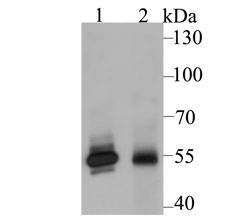
Fig1: Western blot analysis of GLUR on different lysates using anti-GLUR antibody at 1/2,000 dilution. Positive control: Lane1: A549 Lane2: Rat liver tissueApplication
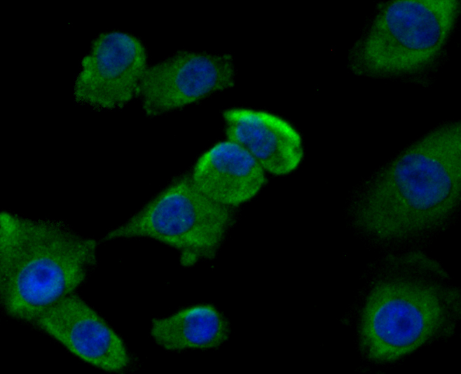
Fig2: ICC staining GLUR in A549 cells (green). The nuclear counter stain is DAPI (blue). Cells were fixed in paraformaldehyde, permeabilised with 0.25% Triton X100/PBS.Application
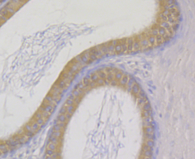
Fig3: Immunohistochemical analysis of paraffin-embedded rat epididymis tissue using anti-GLUR antibody. Counter stained with hematoxylin.Application
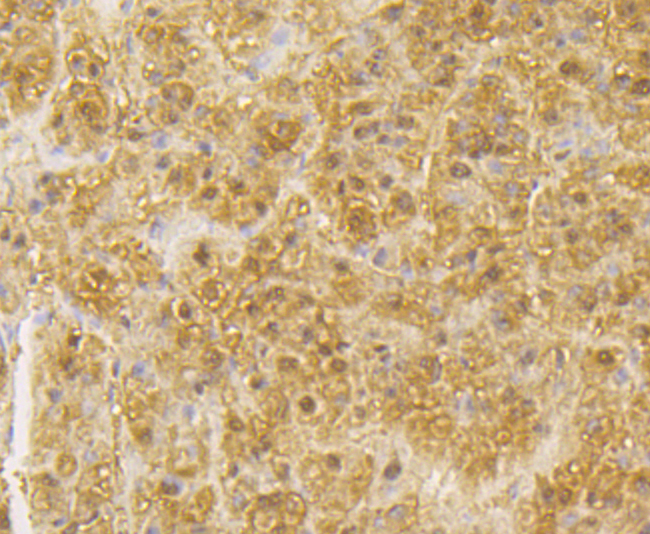
Fig4: Immunohistochemical analysis of paraffin-embedded human liver tissue using anti-GLUR antibody. Counter stained with hematoxylin.Application
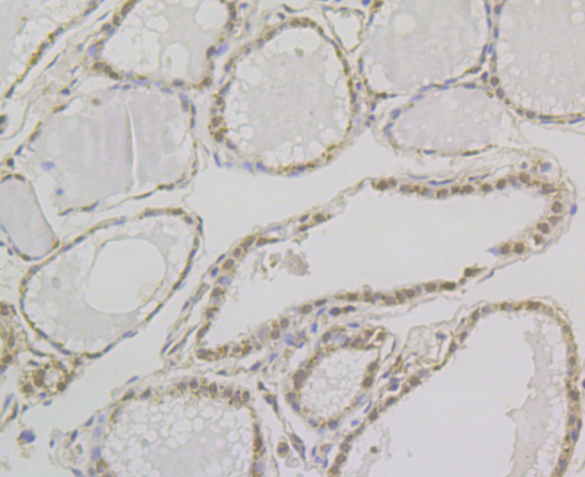
Fig5: Immunohistochemical analysis of paraffin-embedded human thyroid gland tissue using anti-GLUR antibody. Counter stained with hematoxylin.Application
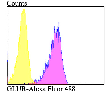
Fig6: Flow cytometric analysis of A549 cells with GLUR antibody at 1/100 dilution (purple) compared with an unlabelled control (cells without incubation with primary antibody; yellow). Alexa Fluor 488-conjugated goat anti-rabbit IgG was used as the secondary antibody.| Product Name | GLUR |
|---|---|
| Antibody Type | Primary Antibodies |
| Antigen Alias | GLR1 antibody GLUR antibody Glutathione reductase antibody Glutathione reductase mitochondrial antibody Glutathione reductase, mitochondrial antibody GR antibody Gr1 antibody GRase antibody GRD 1 antibody GRD1 antibody GSHR_HUMAN antibody GSR antibody MGC78522 antibody |
| Product description | Glutathione plays a key role in maintaining proper function and preventing oxidative stress in human cells. It can act as a scavenger for hydroxyl radicals, singlet oxygen, and various electrophiles. Reduced glutathione reduces the oxidized form of the enzyme glutathione peroxidase, which in turn reduces hydrogen peroxide (H2O2), a dangerously reactive species within the cell. In addition, it plays a key role in the metabolism and clearance of xenobiotics, acts as a cofactor in certain detoxifying enzymes, participates in transport, and regenerates antioxidants such and Vitamins E and C to their reactive forms. The ratio of GSSG/GSH present in the cell is a key factor in properly maintaining the oxidative balance of the cell, that is, it is critical that the cell maintains high levels of the reduced glutathione and a low level of the oxidized Glutathione disulfide. This narrow balance is maintained by glutathione reductase, which catalyzes the reduction of GSSG to GSH. |
| Immunogen | Recombinant protein within human GLUR 260-450 aa. |
| Clonality | Polyclonal |
|---|---|
| Isotype | IgG |
| Host Species | Rabbit |
| Tested Applications | WBICCIHCFC |
| WB:1:1,000-1:2,000 ICC:1:50-1:200 IHC:1:50-1:200 FC:1:50-1:100 |
|
| Species Reactivity | HumanRat |
| Concentration | 1mg/ml |
| Alternative Names | GLR1 antibody
GLUR antibody
Glutathione reductase antibody
Glutathione reductase mitochondrial antibody
Glutathione reductase mitochondrial antibody GR antibody Gr1 antibody GRase antibody GRD 1 antibody GRD1 antibody GSHR_HUMAN antibody GSR antibody MGC78522 antibody |
|---|---|
| Molecular Weight(MW) | 56 kDa |
| Cellular Localization | Cytoplasm. Mitochondrion. |
Application
Fig1: Western blot analysis of GLUR on different lysates using anti-GLUR antibody at 1/2,000 dilution. Positive control: Lane1: A549 Lane2: Rat liver tissue
Application
Fig2: ICC staining GLUR in A549 cells (green). The nuclear counter stain is DAPI (blue). Cells were fixed in paraformaldehyde, permeabilised with 0.25% Triton X100/PBS.
Application
Fig3: Immunohistochemical analysis of paraffin-embedded rat epididymis tissue using anti-GLUR antibody. Counter stained with hematoxylin.
Application
Fig4: Immunohistochemical analysis of paraffin-embedded human liver tissue using anti-GLUR antibody. Counter stained with hematoxylin.
Application
Fig5: Immunohistochemical analysis of paraffin-embedded human thyroid gland tissue using anti-GLUR antibody. Counter stained with hematoxylin.
Application
Fig6: Flow cytometric analysis of A549 cells with GLUR antibody at 1/100 dilution (purple) compared with an unlabelled control (cells without incubation with primary antibody; yellow). Alexa Fluor 488-conjugated goat anti-rabbit IgG was used as the secondary antibody.| Positive Control | A549, rat liver tissue, rat epididymis tissue, human liver tissue, human thyroid gland tissue. |
|---|---|
| Application Notes | WB:1:1,000-1:2,000 ICC:1:50-1:200 IHC:1:50-1:200 FC:1:50-1:100 |
| Form | Liquid |
|---|---|
| Storage Instructions | Store at +4℃ after thawing. Aliquot store at -20℃. Avoid repeated freeze / thaw cycles. |
| Storage Buffer | 1*TBS (pH7.4), 0.5%BSA, 50%Glycerol. Preservative: 0.05% Sodium Azide. |


