Application
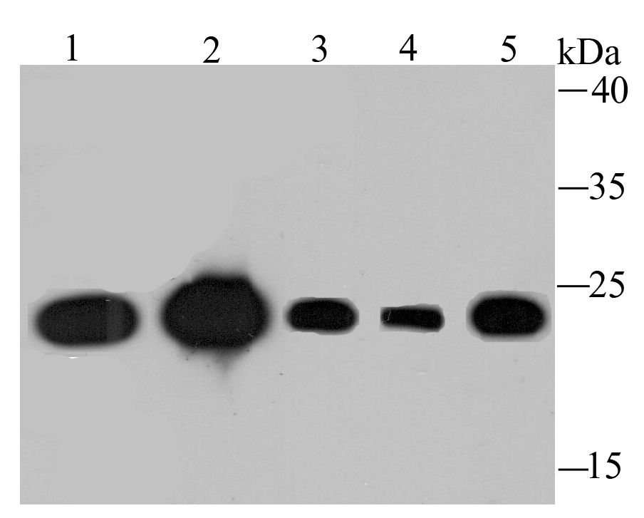
Fig1: Western blot analysis of TCTP on different lysates using anti-TCTP antibody at 1/1,000 dilution. Positive control: Lane 1: Mouse placenta tissue Lane 2: Mouse testis tissue Lane 3: Hela Lane 4: A431 Lane 5: MCF-7Application
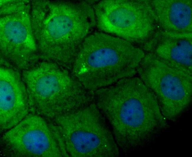
Fig2: ICC staining TCTP in A431 cells (green). The nuclear counter stain is DAPI (blue). Cells were fixed in paraformaldehyde, permeabilised with 0.25% Triton X100/PBS.Application
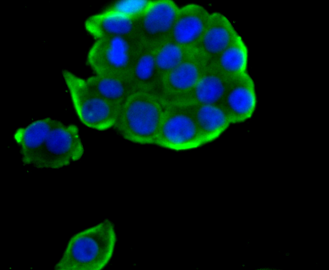
Fig3: ICC staining TCTP in LOVO cells (green). The nuclear counter stain is DAPI (blue). Cells were fixed in paraformaldehyde, permeabilised with 0.25% Triton X100/PBS.Application
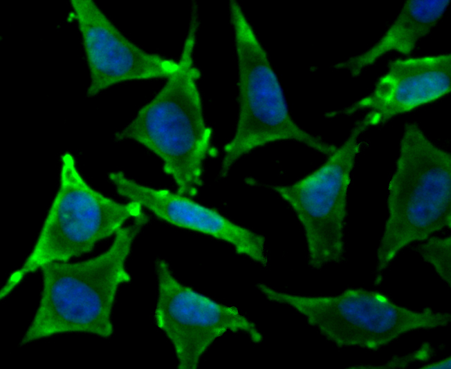
Fig4: ICC staining TCTP in SH-SY5Y cells (green). The nuclear counter stain is DAPI (blue). Cells were fixed in paraformaldehyde, permeabilised with 0.25% Triton X100/PBS.Application
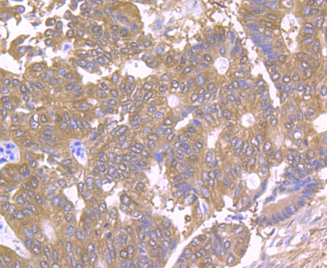
Fig5: Immunohistochemical analysis of paraffin-embedded human colon cancer tissue using anti-TCTP antibody. Counter stained with hematoxylin.Application
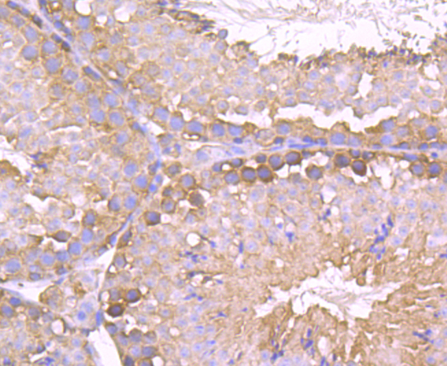
Fig6: Immunohistochemical analysis of paraffin-embedded mouse testis tissue using anti-TCTP antibody. Counter stained with hematoxylin.Application
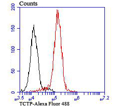
Fig7: Flow cytometric analysis of THP-1 cells with TCTP antibody at 1/100 dilution (red) compared with an unlabelled control (cells without incubation with primary antibody; black). Alexa Fluor® 488-conjugated goat anti-rabbit IgG was used as the secondary antibody.| Product Name | TCTP [JU13-32] |
|---|---|
| Antibody Type | Primary Antibodies |
| Antigen Alias | FLJ27337 antibody Fortilin antibody Histamine releasing factor antibody Histamine-releasing factor antibody HRF antibody Immunoglobulin E-dependent antibody p02 antibody p23 antibody TCTP antibody TCTP_HUMAN antibody tpt1 antibody Translationally controlled tumor protein antibody Translationally-controlled tumor protein antibody Tumor protein translationally controlled 1 antibody tumor protein, translationally-controlled 1 antibody |
| Product description | Histamine-releasing factor (HRF) is a cytokine-like molecule that causes the release of histamine, IL-4 and IL-13 from basophils as well as the secretion of IL-8 and a calcium response in eosinophils. HRF belongs to the translationally controlled tumor protein (TCTP) family. It is expressed in several healthy and tumoral cells, including erythrocytes, hepatocytes, macrophages, platelets, keratinocytes, erythroleukemia cells, gliomas, melanomas, hepatoblastomas and lymphomas, and it is localized in the cytoplasm. HRF plays a pivotal role in allergic diseases, and due to its wide distribution in brain, is thought to be involved in neurodegenerative disorders, such as Alzheimer's disease and Down syndrome. |
| Immunogen | Recombinant protein |
| Clonality | Monoclonal |
|---|---|
| Isotype | IgG |
| Host Species | Recombinant rabbit |
| Tested Applications | WBICC/IFIHCFCIP |
| WB:1:500-1:2,000 ICC/IF:1:50-1:200 IHC:1:50-1:200 FC:1:50-1:100 |
|
| Species Reactivity | HumanMouseRat |
| Concentration | 1mg/ml |
| Alternative Names | FLJ27337 antibody
Fortilin antibody
Histamine releasing factor antibody
Histamine-releasing factor antibody
HRF antibody
Immunoglobulin E-dependent antibody
p02 antibody
p23 antibody
TCTP antibody
TCTP_HUMAN antibody
tpt1 antibody
Translationally controlled tumor protein antibody
Translationally-controlled tumor protein antibody
Tumor protein translationally controlled 1 antibody
tumor protein translationally-controlled 1 antibody |
|---|---|
| Molecular Weight(MW) | 20 kDa |
| Cellular Localization | Cytoplasm. |
Application
Fig1: Western blot analysis of TCTP on different lysates using anti-TCTP antibody at 1/1,000 dilution. Positive control: Lane 1: Mouse placenta tissue Lane 2: Mouse testis tissue Lane 3: Hela Lane 4: A431 Lane 5: MCF-7
Application
Fig2: ICC staining TCTP in A431 cells (green). The nuclear counter stain is DAPI (blue). Cells were fixed in paraformaldehyde, permeabilised with 0.25% Triton X100/PBS.
Application
Fig3: ICC staining TCTP in LOVO cells (green). The nuclear counter stain is DAPI (blue). Cells were fixed in paraformaldehyde, permeabilised with 0.25% Triton X100/PBS.
Application
Fig4: ICC staining TCTP in SH-SY5Y cells (green). The nuclear counter stain is DAPI (blue). Cells were fixed in paraformaldehyde, permeabilised with 0.25% Triton X100/PBS.
Application
Fig5: Immunohistochemical analysis of paraffin-embedded human colon cancer tissue using anti-TCTP antibody. Counter stained with hematoxylin.
Application
Fig6: Immunohistochemical analysis of paraffin-embedded mouse testis tissue using anti-TCTP antibody. Counter stained with hematoxylin.
Application
Fig7: Flow cytometric analysis of THP-1 cells with TCTP antibody at 1/100 dilution (red) compared with an unlabelled control (cells without incubation with primary antibody; black). Alexa Fluor® 488-conjugated goat anti-rabbit IgG was used as the secondary antibody.| Positive Control | Mouse placenta tissue lysate, mouse testis tissue, Hela, A431, MCF-7, LOVO, SH-SY5Y, human colon cancer tissue, THP-1. |
|---|---|
| Application Notes | WB:1:500-1:2,000 ICC/IF:1:50-1:200 IHC:1:50-1:200 FC:1:50-1:100 |
| Form | Liquid |
|---|---|
| Storage Instructions | Store at +4℃ after thawing. Aliquot store at -20℃ or -80℃. Avoid repeated freeze / thaw cycles. |
| Storage Buffer | 1*TBS (pH7.4), 1%BSA, 40%Glycerol. Preservative: 0.05% Sodium Azide. |


