Application
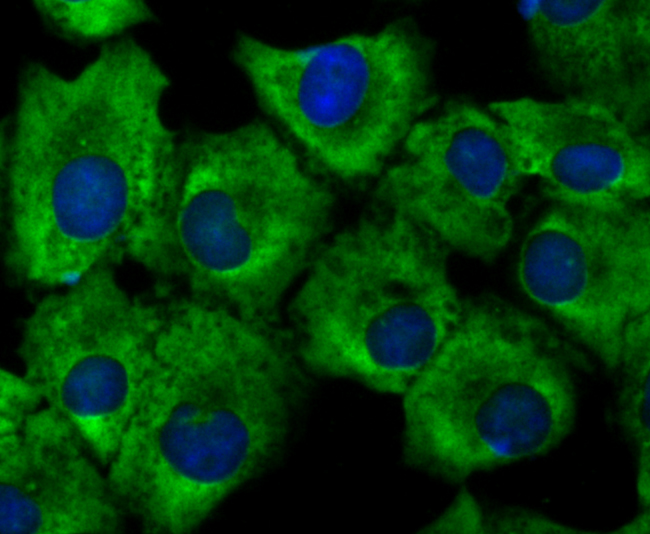
Fig1: ICC staining Growth hormone receptor in A549 cells (green). The nuclear counter stain is DAPI (blue). Cells were fixed in paraformaldehyde, permeabilised with 0.25% Triton X100/PBS.Application
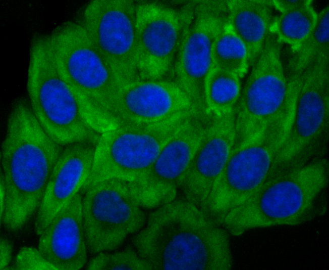
Fig2: ICC staining Growth hormone receptor in HepG2 cells (green). The nuclear counter stain is DAPI (blue). Cells were fixed in paraformaldehyde, permeabilised with 0.25% Triton X100/PBS.Application
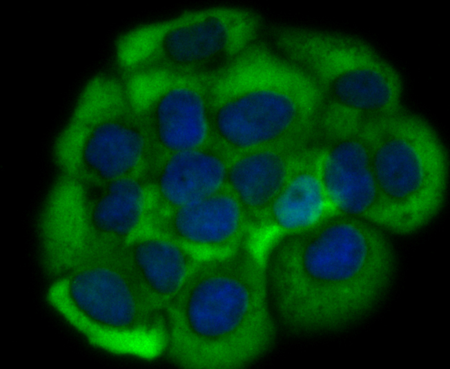
Fig3: ICC staining Growth hormone receptor in JAR cells (green). The nuclear counter stain is DAPI (blue). Cells were fixed in paraformaldehyde, permeabilised with 0.25% Triton X100/PBS.Application
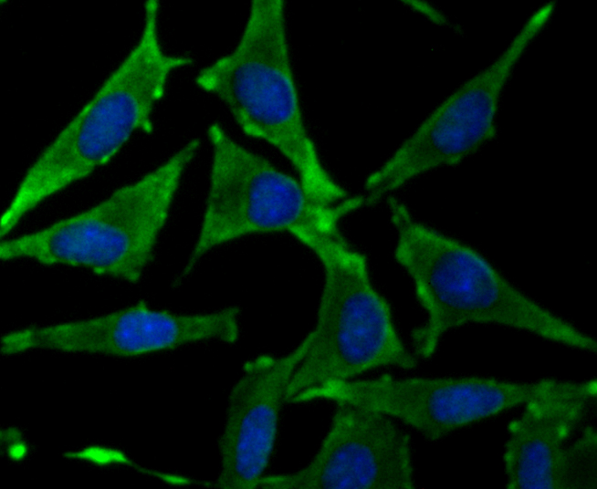
Fig4: ICC staining Growth hormone receptor in SH-SY5Y cells (green). The nuclear counter stain is DAPI (blue). Cells were fixed in paraformaldehyde, permeabilised with 0.25% Triton X100/PBS.Application
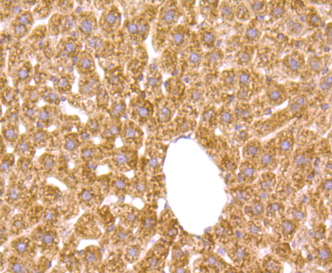
Fig5: Immunohistochemical analysis of paraffin-embedded mouse liver tissue using anti-Growth hormone receptor antibody. Counter stained with hematoxylin.Application
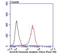
Fig6: Flow cytometric analysis of MCF-7 cells with Growth hormone receptor antibody at 1/100 dilution (red) compared with an unlabelled control (cells without incubation with primary antibody; black). Alexa Fluor 488-conjugated goat anti rabbit IgG was used as the secondary antibody.| Product Name | Growth hormone receptor [JU01-34] |
|---|---|
| Antibody Type | Primary Antibodies |
| Antigen Alias | GH receptor antibody GH-binding protein antibody GHBP antibody GHBP, included antibody GHR antibody GHR_HUMAN antibody Growth hormone binding protein antibody Growth hormone receptor antibody Growth hormone receptor precursor antibody Growth hormone-binding protein antibody Growth hormone-binding protein, included antibody Increased responsiveness to growth hormone, included antibody Serum binding protein antibody Serum-binding protein antibody Somatotropin receptor antibody |
| Product description | GHR (growth hormone receptor) binds growth hormone (GH), which is produced by the anterior pituitary and regulates body growth and other metabolic processes. GHR is an integral membrane protein and a member of the cytokine receptor family. A common characteristic of the cytokine receptor family is having soluble forms of the protein. The soluble form of GHR is GH-binding protein (GHBP), which is generated by the proteolytic cleavage of the extracellular domain of GHR (2-4). Reduced levels of GHBP are associated with GH insensitivity syndrome (GHIS) (2,4). GHR has been shown to be transcribed via at least two different promoters, resulting in GHR 1A and GHR 1B. Both GHR 1A and 1B are expressed in liver, whereas GHR 1B is also expressed in muscle, uterus, and ovary tissues. |
| Immunogen | Recombinant protein |
| Clonality | Monoclonal |
|---|---|
| Isotype | IgG |
| Host Species | Recombinant rabbit |
| Tested Applications | WBICCIHCFC |
| ICC:1:50-1:200 IHC:1:50-1:200 FC:1:50-1:100 WB:1:500 |
|
| Species Reactivity | HumanMouseRat |
| Concentration | 1mg/ml |
| Alternative Names | GH receptor antibody
GH-binding protein antibody
GHBP antibody
GHBP included antibody GHR antibody GHR_HUMAN antibody Growth hormone binding protein antibody Growth hormone receptor antibody Growth hormone receptor precursor antibody Growth hormone-binding protein antibody Growth hormone-binding protein included antibody Increased responsiveness to growth hormone included antibody Serum binding protein antibody Serum-binding protein antibody Somatotropin receptor antibody |
|---|---|
| Molecular Weight(MW) | 71 kDa |
| Cellular Localization | Secreted. Cell membrane. |
Application
Fig1: ICC staining Growth hormone receptor in A549 cells (green). The nuclear counter stain is DAPI (blue). Cells were fixed in paraformaldehyde, permeabilised with 0.25% Triton X100/PBS.
Application
Fig2: ICC staining Growth hormone receptor in HepG2 cells (green). The nuclear counter stain is DAPI (blue). Cells were fixed in paraformaldehyde, permeabilised with 0.25% Triton X100/PBS.
Application
Fig3: ICC staining Growth hormone receptor in JAR cells (green). The nuclear counter stain is DAPI (blue). Cells were fixed in paraformaldehyde, permeabilised with 0.25% Triton X100/PBS.
Application
Fig4: ICC staining Growth hormone receptor in SH-SY5Y cells (green). The nuclear counter stain is DAPI (blue). Cells were fixed in paraformaldehyde, permeabilised with 0.25% Triton X100/PBS.
Application
Fig5: Immunohistochemical analysis of paraffin-embedded mouse liver tissue using anti-Growth hormone receptor antibody. Counter stained with hematoxylin.
Application
Fig6: Flow cytometric analysis of MCF-7 cells with Growth hormone receptor antibody at 1/100 dilution (red) compared with an unlabelled control (cells without incubation with primary antibody; black). Alexa Fluor 488-conjugated goat anti rabbit IgG was used as the secondary antibody.| Positive Control | HepG2, JAR, A549, SH-SY5Y, MCF-7, mouse liver tissue. |
|---|---|
| Application Notes | ICC:1:50-1:200 IHC:1:50-1:200 FC:1:50-1:100 WB:1:500 |
| Form | Liquid |
|---|---|
| Storage Instructions | Store at +4℃ after thawing. Aliquot store at -20℃ or -80℃. Avoid repeated freeze / thaw cycles. |
| Storage Buffer | 1*TBS (pH7.4), 1%BSA, 40%Glycerol. Preservative: 0.05% Sodium Azide. |


