Application
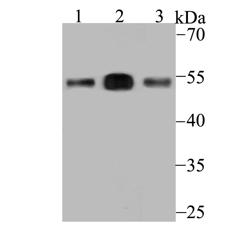
Fig1: Western blot analysis of ERGI3 on different lysates using anti-ERGI3 antibody at 1/500 dilution. Positive control: Lane1: SiHa Lane2: Human placenta Lane3: Rat bone marrowApplication
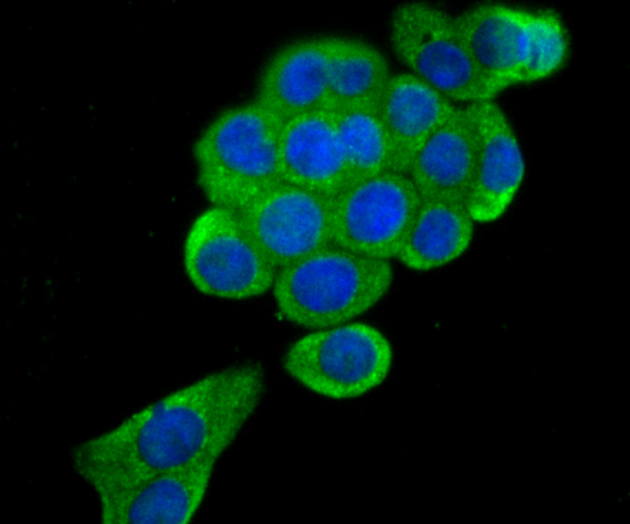
Fig2: ICC staining ERGI3 in LOVO cells (green). The nuclear counter stain is DAPI (blue). Cells were fixed in paraformaldehyde, permeabilised with 0.25% Triton X100/PBS.Application
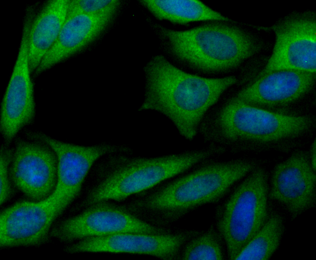
Fig3: ICC staining ERGI3 in SiHa cells (green). The nuclear counter stain is DAPI (blue). Cells were fixed in paraformaldehyde, permeabilised with 0.25% Triton X100/PBS.Application
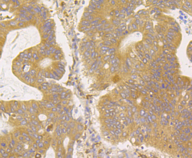
Fig4: Immunohistochemical analysis of paraffin-embedded human colon cancer tissue using anti-ERGI3 antibody. Counter stained with hematoxylin.Application
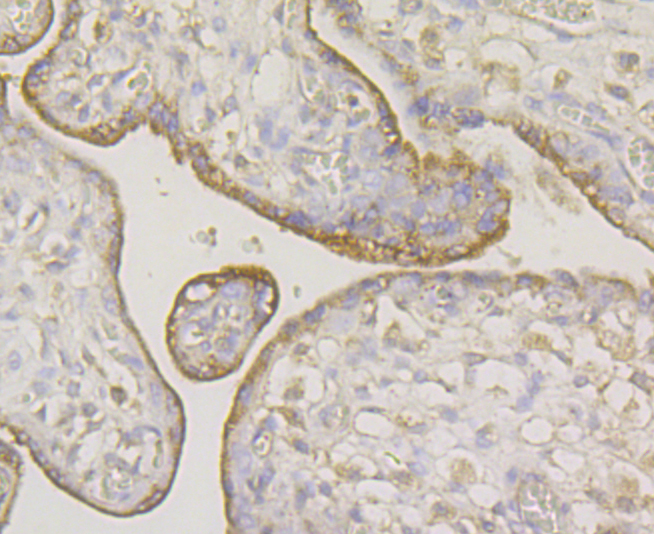
Fig5: Immunohistochemical analysis of paraffin-embedded human placenta tissue using anti-ERGI3 antibody. Counter stained with hematoxylin.Application
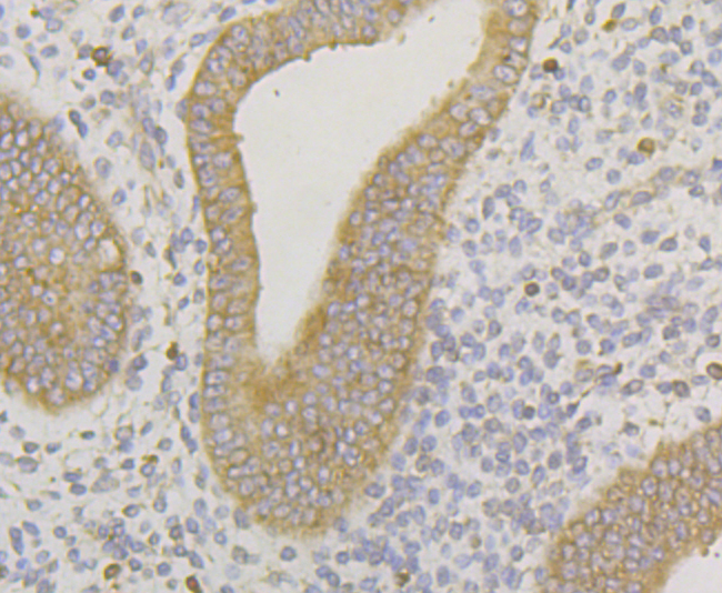
Fig6: Immunohistochemical analysis of paraffin-embedded human uterus tissue using anti-ERGI3 antibody. Counter stained with hematoxylin.Application
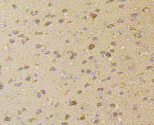
Fig7: Immunohistochemical analysis of paraffin-embedded mouse brain tissue using anti-ERGI3 antibody. Counter stained with hematoxylin.Application
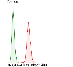
Fig8: Flow cytometric analysis of LOVO cells with ERGI3 antibody at 1/100 dilution (red) compared with an unlabelled control (cells without incubation with primary antibody; green). Alexa Fluor 488-conjugated goat anti-rabbit IgG was used as the secondary antibody.| Product Name | ERGI3 |
|---|---|
| Antibody Type | Primary Antibodies |
| Antigen Alias | 2310015B14Rik antibody AV318804 antibody C20orf47 antibody CGI 54 antibody D2Ucla1 antibody dJ477O4.2 antibody DKFZp547A2190 antibody Endoplasmic reticulum Golgi intermediate compartment protein 3 antibody endoplasmic reticulum localized protein ERp43 antibody Endoplasmic reticulum-Golgi intermediate compartment protein 3 antibody ERGI3_HUMAN antibody ERGIC and golgi 3 antibody ergic3 antibody ERV46 antibody NY BR 84 antibody PRO0989 antibody RP23-220D12.2 antibody RP3-477O4.1 antibody SDBCAG84 antibody Serologically defined breast cancer antigen 84 antibody Serologically defined breast cancer antigen NY BR 84 antibody Serologically defined breast cancer antigen NY-BR-84 antibody |
| Product description | Belongs to the ERGIC family. The ERGIC lies between the rough endoplasmic reticulum and Golgi on the secretory pathway. The vesicular-tubular cluster (VTC), also referred to as the ER-Golgi intermediate compartment (or ERGIC), is an organelle in eukaryotic cells. This compartment mediates trafficking between the endoplasmic reticulum and Golgi complex, facilitating the sorting of cargo. ERGI3 possible role in transport between endoplasmic reticulum and Golgi. |
| Immunogen | Synthetic peptide within C-terminal human ERGI3. |
| Clonality | Polyclonal |
|---|---|
| Isotype | IgG |
| Host Species | Rabbit |
| Tested Applications | WBICCIHCFC |
| WB:1:500 ICC:1:50-1:200 IHC:1:50-1:200 FC:1:50-1:100 |
|
| Species Reactivity | HumanMouseRat |
| Concentration | 1mg/ml |
| Alternative Names | 2310015B14Rik antibody AV318804 antibody C20orf47 antibody CGI 54 antibody D2Ucla1 antibody dJ477O4.2 antibody DKFZp547A2190 antibody Endoplasmic reticulum Golgi intermediate compartment protein 3 antibody endoplasmic reticulum localized protein ERp43 antibody Endoplasmic reticulum-Golgi intermediate compartment protein 3 antibody ERGI3_HUMAN antibody ERGIC and golgi 3 antibody ergic3 antibody ERV46 antibody NY BR 84 antibody PRO0989 antibody RP23-220D12.2 antibody RP3-477O4.1 antibody SDBCAG84 antibody Serologically defined breast cancer antigen 84 antibody Serologically defined breast cancer antigen NY BR 84 antibody Serologically defined breast cancer antigen NY-BR-84 antibody |
|---|---|
| Molecular Weight(MW) | 55 kDa |
| Cellular Localization | Endoplasmic reticulum. Golgi apparatus. |
Application
Fig1: Western blot analysis of ERGI3 on different lysates using anti-ERGI3 antibody at 1/500 dilution. Positive control: Lane1: SiHa Lane2: Human placenta Lane3: Rat bone marrow
Application
Fig2: ICC staining ERGI3 in LOVO cells (green). The nuclear counter stain is DAPI (blue). Cells were fixed in paraformaldehyde, permeabilised with 0.25% Triton X100/PBS.
Application
Fig3: ICC staining ERGI3 in SiHa cells (green). The nuclear counter stain is DAPI (blue). Cells were fixed in paraformaldehyde, permeabilised with 0.25% Triton X100/PBS.
Application
Fig4: Immunohistochemical analysis of paraffin-embedded human colon cancer tissue using anti-ERGI3 antibody. Counter stained with hematoxylin.
Application
Fig5: Immunohistochemical analysis of paraffin-embedded human placenta tissue using anti-ERGI3 antibody. Counter stained with hematoxylin.
Application
Fig6: Immunohistochemical analysis of paraffin-embedded human uterus tissue using anti-ERGI3 antibody. Counter stained with hematoxylin.
Application
Fig7: Immunohistochemical analysis of paraffin-embedded mouse brain tissue using anti-ERGI3 antibody. Counter stained with hematoxylin.
Application
Fig8: Flow cytometric analysis of LOVO cells with ERGI3 antibody at 1/100 dilution (red) compared with an unlabelled control (cells without incubation with primary antibody; green). Alexa Fluor 488-conjugated goat anti-rabbit IgG was used as the secondary antibody.| Positive Control | Siha, human placenta tissue, rat bone marrow tissue, LOVO, human colon cancer tissue, human uterus tissue, mouse brain tissue. |
|---|---|
| Application Notes | WB:1:500 ICC:1:50-1:200 IHC:1:50-1:200 FC:1:50-1:100 |
| Form | Liquid |
|---|---|
| Storage Instructions | Store at +4℃ after thawing. Aliquot store at -20℃. Avoid repeated freeze / thaw cycles. |
| Storage Buffer | 1*TBS (pH7.4), 0.5%BSA, 50%Glycerol. Preservative: 0.05% Sodium Azide. |


