Application
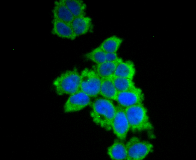
Fig1: ICC staining UAP1 in 293T cells (green). The nuclear counter stain is DAPI (blue). Cells were fixed in paraformaldehyde, permeabilised with 0.25% Triton X100/PBS.Application
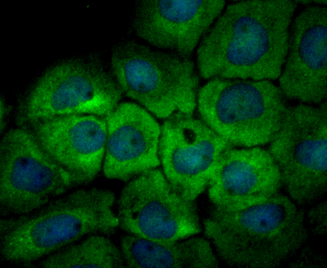
Fig2: ICC staining UAP1 in A431 cells (green). The nuclear counter stain is DAPI (blue). Cells were fixed in paraformaldehyde, permeabilised with 0.25% Triton X100/PBS.Application
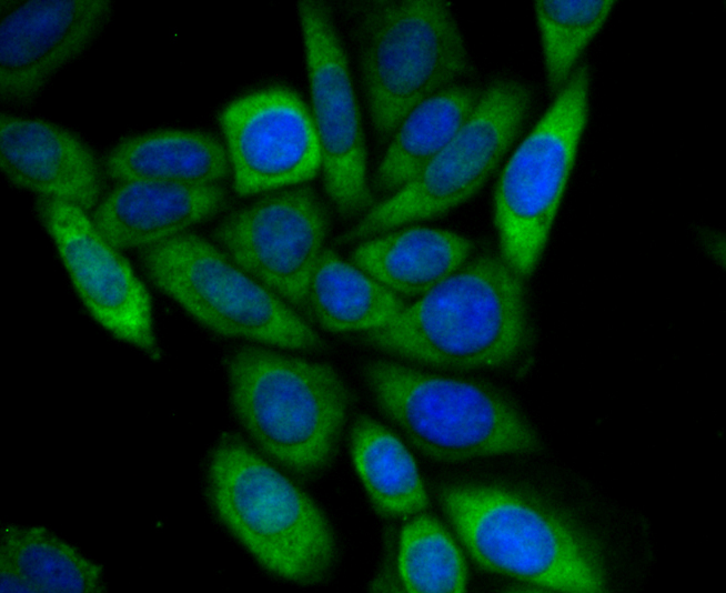
Fig3: ICC staining UAP1 in SiHa cells (green). The nuclear counter stain is DAPI (blue). Cells were fixed in paraformaldehyde, permeabilised with 0.25% Triton X100/PBS.Application
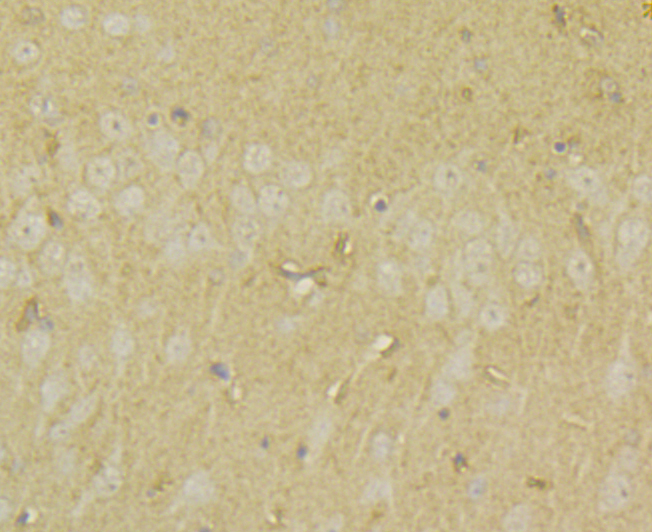
Fig4: Immunohistochemical analysis of paraffin-embedded rat brain tissue using anti-UAP1 antibody. Counter stained with hematoxylin.Application
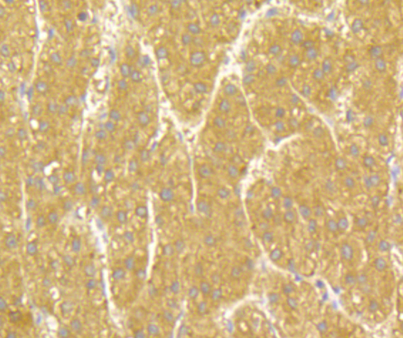
Fig5: Immunohistochemical analysis of paraffin-embedded human liver tissue using anti-UAP1 antibody. Counter stained with hematoxylin.Application
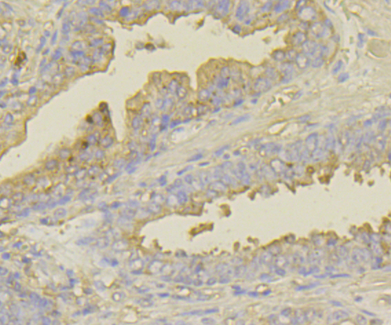
Fig6: Immunohistochemical analysis of paraffin-embedded human prostate cancer tissue using anti-UAP1 antibody. Counter stained with hematoxylin.Application
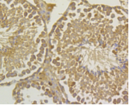
Fig7: Immunohistochemical analysis of paraffin-embedded mouse testis tissue using anti-UAP1 antibody. Counter stained with hematoxylin.Application
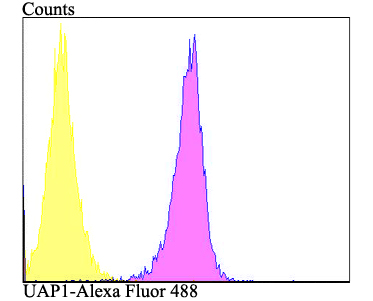
Fig8: Flow cytometric analysis of K562 cells with UAP1 antibody at 1/100 dilution (purple) compared with an unlabelled control (cells without incubation with primary antibody; yellow). Alexa Fluor 488-conjugated goat anti-rabbit IgG was used as the secondary antibody.| Product Name | UAP1 |
|---|---|
| Antibody Type | Primary Antibodies |
| Antigen Alias | AGX 1 antibody AGX antibody AGX-1 antibody AGX-2 antibody AGX1 antibody Antigen X antibody AntigenX antibody SPAG 2 antibody SPAG2 antibody Sperm associated antigen 2 antibody Sperm-associated antigen 2 antibody UAP 1 antibody uap1 antibody UAP1_HUMAN antibody UDP N acetylhexosamine pyrophosphorylase antibody UDP N acteylglucosamine pyrophosphorylase 1 antibody UDP-N-acetylglucosamine pyrophosphorylase antibody |
| Product description | Converts UDP and GlcNAc-1-P into UDP-GlcNAc, and UDP and GalNAc-1-P into UDP-GalNAc. Isoform AGX1 has 2 to 3 times higher activity towards GalNAc-1-P, while isoform AGX2 has 8 times more activity towards GlcNAc-1-P. |
| Immunogen | Synthetic peptide within human UAP1 20-100aa. |
| Clonality | Polyclonal |
|---|---|
| Isotype | IgG |
| Host Species | Rabbit |
| Tested Applications | ICCIHCFC |
| ICC:1:50-1:200 IHC:1:50-1:200 FC:1:50-1:100 |
|
| Species Reactivity | HumanMouseRat |
| Concentration | 1mg/ml |
| Alternative Names | AGX 1 antibody AGX antibody AGX-1 antibody AGX-2 antibody AGX1 antibody Antigen X antibody AntigenX antibody SPAG 2 antibody SPAG2 antibody Sperm associated antigen 2 antibody Sperm-associated antigen 2 antibody UAP 1 antibody uap1 antibody UAP1_HUMAN antibody UDP N acetylhexosamine pyrophosphorylase antibody UDP N acteylglucosamine pyrophosphorylase 1 antibody UDP-N-acetylglucosamine pyrophosphorylase antibody |
|---|---|
| Molecular Weight(MW) | 58 kDa |
| Cellular Localization | Cytoplasm. |
Application
Fig1: ICC staining UAP1 in 293T cells (green). The nuclear counter stain is DAPI (blue). Cells were fixed in paraformaldehyde, permeabilised with 0.25% Triton X100/PBS.
Application
Fig2: ICC staining UAP1 in A431 cells (green). The nuclear counter stain is DAPI (blue). Cells were fixed in paraformaldehyde, permeabilised with 0.25% Triton X100/PBS.
Application
Fig3: ICC staining UAP1 in SiHa cells (green). The nuclear counter stain is DAPI (blue). Cells were fixed in paraformaldehyde, permeabilised with 0.25% Triton X100/PBS.
Application
Fig4: Immunohistochemical analysis of paraffin-embedded rat brain tissue using anti-UAP1 antibody. Counter stained with hematoxylin.
Application
Fig5: Immunohistochemical analysis of paraffin-embedded human liver tissue using anti-UAP1 antibody. Counter stained with hematoxylin.
Application
Fig6: Immunohistochemical analysis of paraffin-embedded human prostate cancer tissue using anti-UAP1 antibody. Counter stained with hematoxylin.
Application
Fig7: Immunohistochemical analysis of paraffin-embedded mouse testis tissue using anti-UAP1 antibody. Counter stained with hematoxylin.
Application
Fig8: Flow cytometric analysis of K562 cells with UAP1 antibody at 1/100 dilution (purple) compared with an unlabelled control (cells without incubation with primary antibody; yellow). Alexa Fluor 488-conjugated goat anti-rabbit IgG was used as the secondary antibody.| Positive Control | 293T, A431, SiHa, rat brain tissue, human liver tissue, human prostate cancer tissue, mouse testis tissue, K562. |
|---|---|
| Application Notes | ICC:1:50-1:200 IHC:1:50-1:200 FC:1:50-1:100 |
| Form | Liquid |
|---|---|
| Storage Instructions | Store at +4℃ after thawing. Aliquot store at -20℃. Avoid repeated freeze / thaw cycles. |
| Storage Buffer | 1*TBS (pH7.4), 0.5%BSA, 50%Glycerol. Preservative: 0.05% Sodium Azide. |


