Application
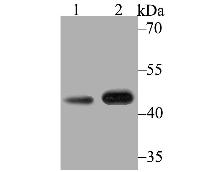
Fig1: Western blot analysis of Kir3.4 on different lysates using anti-Kir3.4 antibody at 1/500 dilution. Positive control: Lane1: Mouse heart Lane2: Rat heartApplication
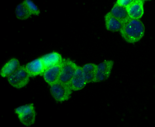
Fig2: ICC staining Kir3.4 in 293T cells (green). The nuclear counter stain is DAPI (blue). Cells were fixed in paraformaldehyde, permeabilised with 0.25% Triton X100/PBS.Application
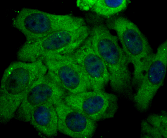
Fig3: ICC staining Kir3.4 in HepG2 cells (green). The nuclear counter stain is DAPI (blue). Cells were fixed in paraformaldehyde, permeabilised with 0.25% Triton X100/PBS.Application
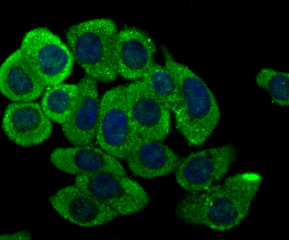
Fig4: ICC staining Kir3.4 in LOVO cells (green). The nuclear counter stain is DAPI (blue). Cells were fixed in paraformaldehyde, permeabilised with 0.25% Triton X100/PBS.Application
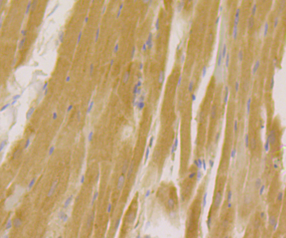
Fig5: Immunohistochemical analysis of paraffin-embedded rat heart tissue using anti-Kir3.4 antibody. Counter stained with hematoxylin.Application
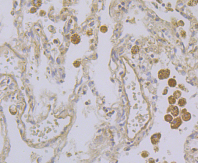
Fig6: Immunohistochemical analysis of paraffin-embedded human lung cancer tissue using anti-Kir3.4 antibody. Counter stained with hematoxylin.Application
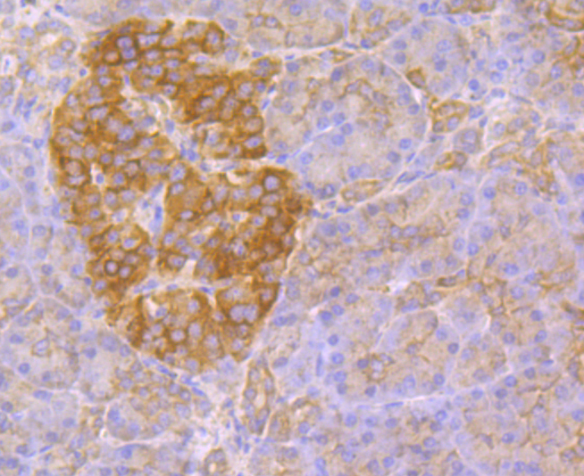
Fig7: Immunohistochemical analysis of paraffin-embedded human pancreas tissue using anti-Kir3.4 antibody. Counter stained with hematoxylin.Application
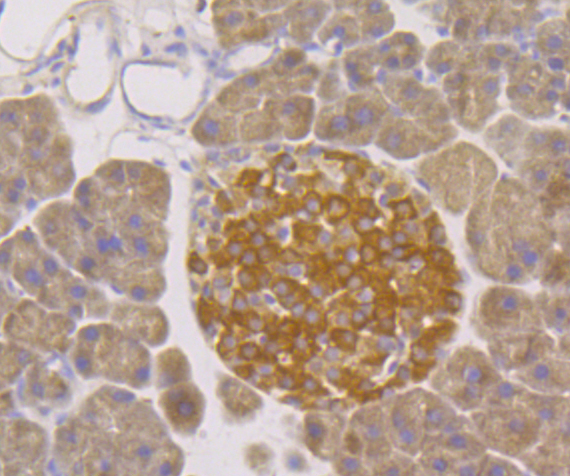
Fig8: Immunohistochemical analysis of paraffin-embedded mouse pancreas tissue using anti-Kir3.4 antibody. Counter stained with hematoxylin.Application
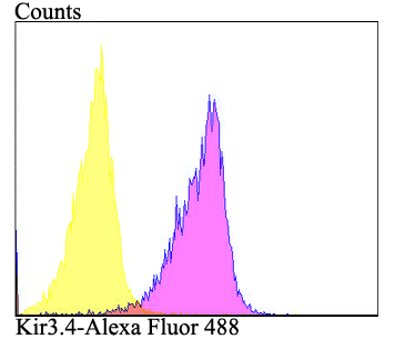
Fig9: Flow cytometric analysis of HepG2 cells with Kir3.4 antibody at 1/100 dilution (purple) compared with an unlabelled control (cells without incubation with primary antibody; yellow). Alexa Fluor 488-conjugated goat anti-rabbit IgG was used as the secondary antibody.| Product Name | Kir3.4 |
|---|---|
| Antibody Type | Primary Antibodies |
| Antigen Alias | Cardiac ATP sensitive potassium channel antibody Cardiac inward rectifier antibody CIR antibody G protein activated inward rectifier potassium channel 4 antibody G protein-activated inward rectifier potassium channel 4 antibody GIRK 4 antibody GIRK-4 antibody GIRK4 antibody Heart KATP channel antibody Inward rectifier K(+) channel Kir3.4 antibody Inward rectifier K+ channel KIR3.4 antibody Inward rectifier potassium channel KIR3.4 antibody inwardly rectifying subfamily J member 5 antibody IRK-4 antibody IRK5_HUMAN antibody KATP 1 antibody KATP-1 antibody KATP1 antibody KCNJ 5 antibody Kcnj5 antibody KIR 3.4 antibody KIR3.4 antibody LQT13 antibody Potassium channel antibody Potassium channel inwardly rectifying subfamily J member 5 antibody Potassium inwardly rectifying channel J5 antibody Potassium inwardly rectifying channel subfamily J member 5 antibody potassium voltage-gated channel subfamily J member 5 antibody |
| Product description | This potassium channel is controlled by G proteins. Inward rectifier potassium channels are characterized by a greater tendency to allow potassium to flow into the cell rather than out of it. Their voltage dependence is regulated by the concentration of extracellular potassium; as external potassium is raised, the voltage range of the channel opening shifts to more positive voltages. The inward rectification is mainly due to the blockage of outward current by internal magnesium. Can be blocked by external barium. |
| Immunogen | Synthetic peptide within N-terminal human Kir3.4. |
| Clonality | Polyclonal |
|---|---|
| Isotype | IgG |
| Host Species | Rabbit |
| Tested Applications | WBICCIHCFC |
| WB:1:500 ICC:1:50-1:200 IHC:1:50-1:200 FC:1:50-1:100 |
|
| Species Reactivity | HumanMouseRat |
| Concentration | 1mg/ml |
| Alternative Names | Cardiac ATP sensitive potassium channel antibody Cardiac inward rectifier antibody CIR antibody G protein activated inward rectifier potassium channel 4 antibody G protein-activated inward rectifier potassium channel 4 antibody GIRK 4 antibody GIRK-4 antibody GIRK4 antibody Heart KATP channel antibody Inward rectifier K(+) channel Kir3.4 antibody Inward rectifier K+ channel KIR3.4 antibody Inward rectifier potassium channel KIR3.4 antibody inwardly rectifying subfamily J member 5 antibody IRK-4 antibody IRK5_HUMAN antibody KATP 1 antibody KATP-1 antibody KATP1 antibody KCNJ 5 antibody Kcnj5 antibody KIR 3.4 antibody KIR3.4 antibody LQT13 antibody Potassium channel antibody Potassium channel inwardly rectifying subfamily J member 5 antibody Potassium inwardly rectifying channel J5 antibody Potassium inwardly rectifying channel subfamily J member 5 antibody potassium voltage-gated channel subfamily J member 5 antibody |
|---|---|
| Molecular Weight(MW) | 48 kDa |
| Cellular Localization | Membrane. |
Application
Fig1: Western blot analysis of Kir3.4 on different lysates using anti-Kir3.4 antibody at 1/500 dilution. Positive control: Lane1: Mouse heart Lane2: Rat heart
Application
Fig2: ICC staining Kir3.4 in 293T cells (green). The nuclear counter stain is DAPI (blue). Cells were fixed in paraformaldehyde, permeabilised with 0.25% Triton X100/PBS.
Application
Fig3: ICC staining Kir3.4 in HepG2 cells (green). The nuclear counter stain is DAPI (blue). Cells were fixed in paraformaldehyde, permeabilised with 0.25% Triton X100/PBS.
Application
Fig4: ICC staining Kir3.4 in LOVO cells (green). The nuclear counter stain is DAPI (blue). Cells were fixed in paraformaldehyde, permeabilised with 0.25% Triton X100/PBS.
Application
Fig5: Immunohistochemical analysis of paraffin-embedded rat heart tissue using anti-Kir3.4 antibody. Counter stained with hematoxylin.
Application
Fig6: Immunohistochemical analysis of paraffin-embedded human lung cancer tissue using anti-Kir3.4 antibody. Counter stained with hematoxylin.
Application
Fig7: Immunohistochemical analysis of paraffin-embedded human pancreas tissue using anti-Kir3.4 antibody. Counter stained with hematoxylin.
Application
Fig8: Immunohistochemical analysis of paraffin-embedded mouse pancreas tissue using anti-Kir3.4 antibody. Counter stained with hematoxylin.
Application
Fig9: Flow cytometric analysis of HepG2 cells with Kir3.4 antibody at 1/100 dilution (purple) compared with an unlabelled control (cells without incubation with primary antibody; yellow). Alexa Fluor 488-conjugated goat anti-rabbit IgG was used as the secondary antibody.| Positive Control | Mouse heart tissue, rat heart tissue, 293T, HepG2, LOVO, human lung cancer tissue, human pancreas tissue, mouse pancreas tissue. |
|---|---|
| Application Notes | WB:1:500 ICC:1:50-1:200 IHC:1:50-1:200 FC:1:50-1:100 |
| Form | Liquid |
|---|---|
| Storage Instructions | Store at +4℃ after thawing. Aliquot store at -20℃. Avoid repeated freeze / thaw cycles. |
| Storage Buffer | 1*TBS (pH7.4), 0.5%BSA, 50%Glycerol. Preservative: 0.05% Sodium Azide. |


