Application
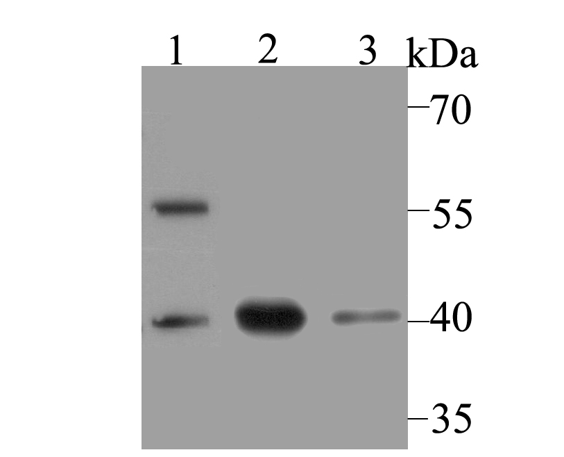
Fig1: Western blot analysis of macroH2A.1 on different lysates using anti-macroH2A.1 antibody at 1/500 dilution. Positive control: Lane1: mouse pancreas Lane2: SiHa Lane3: PC-3MApplication
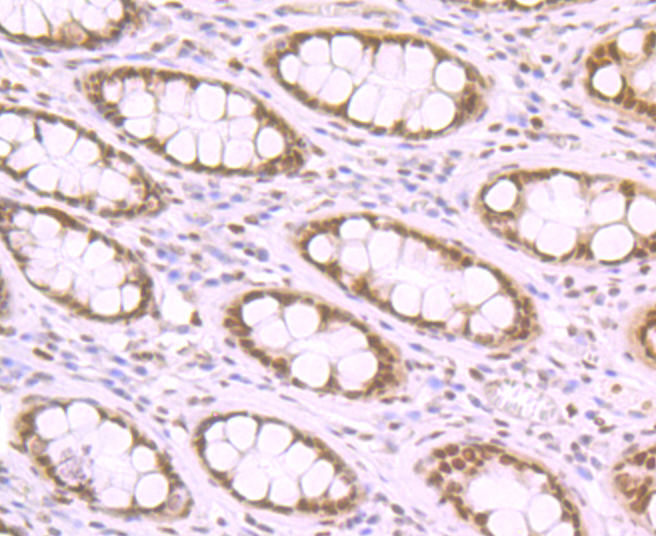
Fig2: Immunohistochemical analysis of paraffin-embedded human colon tissue using anti-macroH2A.1 antibody. Counter stained with hematoxylin.Application
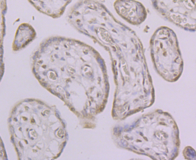
Fig3: Immunohistochemical analysis of paraffin-embedded human placenta tissue using anti-macroH2A.1 antibody. Counter stained with hematoxylin.Application
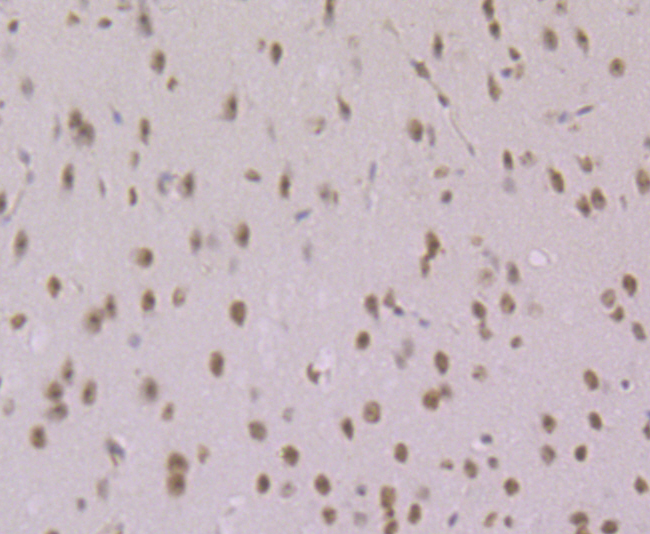
Fig4: Immunohistochemical analysis of paraffin-embedded mouse brain tissue using anti-macroH2A.1 antibody. Counter stained with hematoxylin.Application
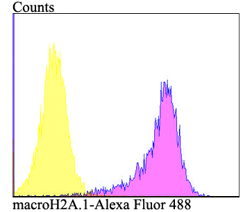
Fig5: Flow cytometric analysis of SiHa cells with macroH2A.1 antibody at 1/100 dilution (purple) compared with an unlabelled control (cells without incubation with primary antibody; yellow). Alexa Fluor 488-conjugated goat anti-rabbit IgG was used as the secondary antibody.| Product Name | macroH2A.1 |
|---|---|
| Antibody Type | Primary Antibodies |
| Antigen Alias | Core histone macro h2a.1 antibody Core histone macro-H2A.1 antibody H2A histone family member Y antibody H2A.y antibody H2A/y antibody H2AF12M antibody H2AFJ antibody H2afy antibody H2AY_HUMAN antibody Histone H2A.Y antibody Histone macroH2A1 antibody Histone macroH2A1.1 antibody Histone macroH2A1.2 antibody Macroh2a1 antibody MACROH2A1.1 antibody MacroH2A1.2 antibody Medulloblastoma antigen MU MB 50.205 antibody Medulloblastoma antigen MU-MB-50.205 antibody mH2a antibody mH2A1 antibody |
| Product description | Variant histone H2A which replaces conventional H2A in a subset of nucleosomes where it represses transcription. Nucleosomes wrap and compact DNA into chromatin, limiting DNA accessibility to the cellular machineries which require DNA as a template. Histones thereby play a central role in transcription regulation, DNA repair, DNA replication and chromosomal stability. DNA accessibility is regulated via a complex set of post-translational modifications of histones, also called histone code, and nucleosome remodeling. Involved in stable X chromosome inactivation. Inhibits the binding of transcription factors, including NF-kappa-B, and interferes with the activity of remodeling SWI/SNF complexes. Inhibits histone acetylation by EP300 and recruits class I HDACs, which induces a hypoacetylated state of chromatin. |
| Immunogen | Recombinant protein within human macroH2A.1 aa 200-400. |
| Clonality | Polyclonal |
|---|---|
| Isotype | IgG |
| Host Species | Rabbit |
| Tested Applications | WBIHCFC |
| WB:1:500 IHC:1:50-1:200 FC:1:50-1:100 |
|
| Species Reactivity | HumanMouse |
| Concentration | 1mg/ml |
| Alternative Names | Core histone macro h2a.1 antibody Core histone macro-H2A.1 antibody H2A histone family member Y antibody H2A.y antibody H2A/y antibody H2AF12M antibody H2AFJ antibody H2afy antibody H2AY_HUMAN antibody Histone H2A.Y antibody Histone macroH2A1 antibody Histone macroH2A1.1 antibody Histone macroH2A1.2 antibody Macroh2a1 antibody MACROH2A1.1 antibody MacroH2A1.2 antibody Medulloblastoma antigen MU MB 50.205 antibody Medulloblastoma antigen MU-MB-50.205 antibody mH2a antibody mH2A1 antibody |
|---|---|
| Molecular Weight(MW) | 40 kDa |
| Cellular Localization | Nucleus. |
Application
Fig1: Western blot analysis of macroH2A.1 on different lysates using anti-macroH2A.1 antibody at 1/500 dilution. Positive control: Lane1: mouse pancreas Lane2: SiHa Lane3: PC-3M
Application
Fig2: Immunohistochemical analysis of paraffin-embedded human colon tissue using anti-macroH2A.1 antibody. Counter stained with hematoxylin.
Application
Fig3: Immunohistochemical analysis of paraffin-embedded human placenta tissue using anti-macroH2A.1 antibody. Counter stained with hematoxylin.
Application
Fig4: Immunohistochemical analysis of paraffin-embedded mouse brain tissue using anti-macroH2A.1 antibody. Counter stained with hematoxylin.
Application
Fig5: Flow cytometric analysis of SiHa cells with macroH2A.1 antibody at 1/100 dilution (purple) compared with an unlabelled control (cells without incubation with primary antibody; yellow). Alexa Fluor 488-conjugated goat anti-rabbit IgG was used as the secondary antibody.| Positive Control | Mouse pancreas tissue, SiHa, PC-3M, human colon tissue, human placenta tissue, mouse brain tissue. |
|---|---|
| Application Notes | WB:1:500 IHC:1:50-1:200 FC:1:50-1:100 |
| Form | Liquid |
|---|---|
| Storage Instructions | Store at +4℃ after thawing. Aliquot store at -20℃. Avoid repeated freeze / thaw cycles. |
| Storage Buffer | 1*TBS (pH7.4), 0.5%BSA, 50%Glycerol. Preservative: 0.05% Sodium Azide. |


