Application
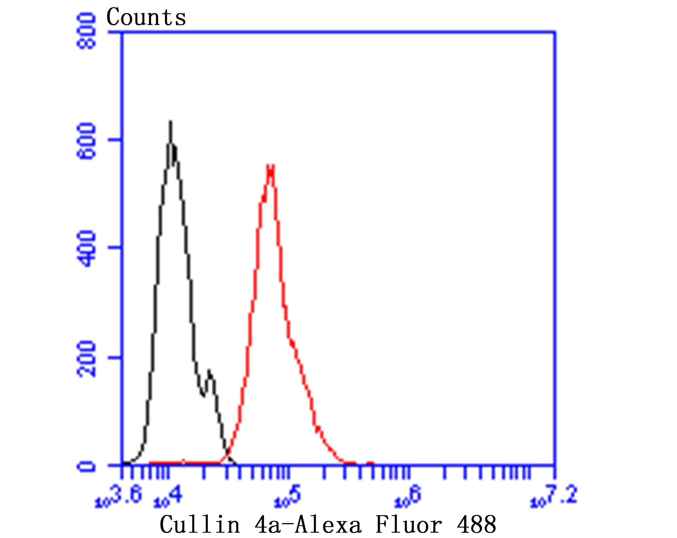
Fig9: Flow cytometric analysis of Hela cells with Cullin 4a antibody at 1/100 dilution (green) compared with an unlabelled control (cells without incubation with primary antibody; red). Alexa Fluor 488-conjugated goat anti-rabbit IgG was used as the secondary antibody.Application
Fig9: Flow cytometric analysis of Hela cells with Cullin 4a antibody at 1/100 dilution (green) compared with an unlabelled control (cells without incubation with primary antibody; red). Alexa Fluor 488-conjugated goat anti-rabbit IgG was used as the secondary antibody.Application
Fig9: Flow cytometric analysis of Hela cells with Cullin 4a antibody at 1/100 dilution (green) compared with an unlabelled control (cells without incubation with primary antibody; red). Alexa Fluor 488-conjugated goat anti-rabbit IgG was used as the secondary antibody.Application
Fig9: Flow cytometric analysis of Hela cells with Cullin 4a antibody at 1/100 dilution (green) compared with an unlabelled control (cells without incubation with primary antibody; red). Alexa Fluor 488-conjugated goat anti-rabbit IgG was used as the secondary antibody.Application
Fig9: Flow cytometric analysis of Hela cells with Cullin 4a antibody at 1/100 dilution (green) compared with an unlabelled control (cells without incubation with primary antibody; red). Alexa Fluor 488-conjugated goat anti-rabbit IgG was used as the secondary antibody.Application
Fig9: Flow cytometric analysis of Hela cells with Cullin 4a antibody at 1/100 dilution (green) compared with an unlabelled control (cells without incubation with primary antibody; red). Alexa Fluor 488-conjugated goat anti-rabbit IgG was used as the secondary antibody.Application
Fig9: Flow cytometric analysis of Hela cells with Cullin 4a antibody at 1/100 dilution (green) compared with an unlabelled control (cells without incubation with primary antibody; red). Alexa Fluor 488-conjugated goat anti-rabbit IgG was used as the secondary antibody.Application
Fig9: Flow cytometric analysis of Hela cells with Cullin 4a antibody at 1/100 dilution (green) compared with an unlabelled control (cells without incubation with primary antibody; red). Alexa Fluor 488-conjugated goat anti-rabbit IgG was used as the secondary antibody.Application
Fig9: Flow cytometric analysis of Hela cells with Cullin 4a antibody at 1/100 dilution (green) compared with an unlabelled control (cells without incubation with primary antibody; red). Alexa Fluor 488-conjugated goat anti-rabbit IgG was used as the secondary antibody.| Product Name | Cullin 4a [JU07-33] |
|---|---|
| Antibody Type | Primary Antibodies |
| Antigen Alias | 2810470J21Rik antibody AW495282 antibody CUL 4A antibody CUL-4A antibody Cul4a antibody Cul4a protein antibody CUL4A_HUMAN antibody Cullin-4A antibody MGC36573 antibody MGC64071 antibody |
| Product description | Core component of multiple cullin-RING-based E3 ubiquitin-protein ligase complexes which mediate the ubiquitination of target proteins. As a scaffold protein may contribute to catalysis through positioning of the substrate and the ubiquitin-conjugating enzyme. The E3 ubiquitin-protein ligase activity of the complex is dependent on the neddylation of the cullin subunit and is inhibited by the association of the deneddylated cullin subunit with TIP120A/CAND1. The functional specificity of the E3 ubiquitin-protein ligase complex depends on the variable substrate recognition component. DCX(DET1-COP1) directs ubiquitination of JUN. DCX(DDB2) directs ubiquitination of XPC. DCX(DDB2) ubiquitinates histones H3-H4 and is required for efficient histone deposition during replication-coupled (H3.1) and replication-independent (H3.3) nucleosome assembly, probably by facilitating the transfer of H3 from ASF1A/ASF1B to other chaperones involved in histone deposition. |
| Immunogen | Recombinant protein |
| Clonality | Monoclonal |
|---|---|
| Isotype | IgG |
| Host Species | Recombinant rabbit |
| Tested Applications | WBICCIHCFC |
| WB:1:500-2,000 ICC:1:50-1:200 IHC:1:50-1:200 FC:1:50-1:100 |
|
| Species Reactivity | Human |
| Concentration | 1mg/ml |
| Alternative Names | 2810470J21Rik antibody AW495282 antibody CUL 4A antibody CUL-4A antibody Cul4a antibody Cul4a protein antibody CUL4A_HUMAN antibody Cullin-4A antibody MGC36573 antibody MGC64071 antibody |
|---|---|
| Molecular Weight(MW) | 88 kDa |
| Cellular Localization | Nucleus. |
| SwissProt ID | Q13619 |
|---|
Application
Fig9: Flow cytometric analysis of Hela cells with Cullin 4a antibody at 1/100 dilution (green) compared with an unlabelled control (cells without incubation with primary antibody; red). Alexa Fluor 488-conjugated goat anti-rabbit IgG was used as the secondary antibody.
Application
Fig9: Flow cytometric analysis of Hela cells with Cullin 4a antibody at 1/100 dilution (green) compared with an unlabelled control (cells without incubation with primary antibody; red). Alexa Fluor 488-conjugated goat anti-rabbit IgG was used as the secondary antibody.
Application
Fig9: Flow cytometric analysis of Hela cells with Cullin 4a antibody at 1/100 dilution (green) compared with an unlabelled control (cells without incubation with primary antibody; red). Alexa Fluor 488-conjugated goat anti-rabbit IgG was used as the secondary antibody.
Application
Fig9: Flow cytometric analysis of Hela cells with Cullin 4a antibody at 1/100 dilution (green) compared with an unlabelled control (cells without incubation with primary antibody; red). Alexa Fluor 488-conjugated goat anti-rabbit IgG was used as the secondary antibody.
Application
Fig9: Flow cytometric analysis of Hela cells with Cullin 4a antibody at 1/100 dilution (green) compared with an unlabelled control (cells without incubation with primary antibody; red). Alexa Fluor 488-conjugated goat anti-rabbit IgG was used as the secondary antibody.
Application
Fig9: Flow cytometric analysis of Hela cells with Cullin 4a antibody at 1/100 dilution (green) compared with an unlabelled control (cells without incubation with primary antibody; red). Alexa Fluor 488-conjugated goat anti-rabbit IgG was used as the secondary antibody.
Application
Fig9: Flow cytometric analysis of Hela cells with Cullin 4a antibody at 1/100 dilution (green) compared with an unlabelled control (cells without incubation with primary antibody; red). Alexa Fluor 488-conjugated goat anti-rabbit IgG was used as the secondary antibody.
Application
Fig9: Flow cytometric analysis of Hela cells with Cullin 4a antibody at 1/100 dilution (green) compared with an unlabelled control (cells without incubation with primary antibody; red). Alexa Fluor 488-conjugated goat anti-rabbit IgG was used as the secondary antibody.
Application
Fig9: Flow cytometric analysis of Hela cells with Cullin 4a antibody at 1/100 dilution (green) compared with an unlabelled control (cells without incubation with primary antibody; red). Alexa Fluor 488-conjugated goat anti-rabbit IgG was used as the secondary antibody.| Positive Control | Hela, A431, HUVEC, human liver cance tissue, human breast cancer tissue, human colon cancer tissue, human kidney tissue, human cervix tissue. |
|---|---|
| Application Notes | WB:1:500-2,000 ICC:1:50-1:200 IHC:1:50-1:200 FC:1:50-1:100 |
| Form | Liquid |
|---|---|
| Storage Instructions | Store at +4℃ after thawing. Aliquot store at -20℃ or -80℃. Avoid repeated freeze / thaw cycles. |
| Storage Buffer | 1*TBS (pH7.4), 1%BSA, 40%Glycerol. Preservative: 0.05% Sodium Azide. |


