Application
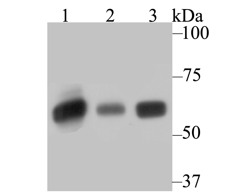
Fig1: Western blot analysis of SERPINC1 on different lysates using anti-SERPINC1 antibody at 1/500 dilution. Positive control: Lane1: HepG2 Lane2: Mouse smooth muscle Lane3: Mouse lungApplication
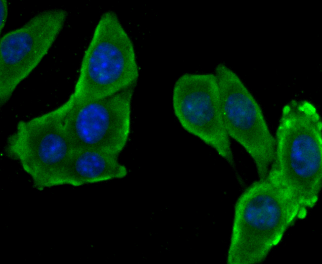
Fig2: ICC staining SERPINC1 in H22 cells (green). The nuclear counter stain is DAPI (blue). Cells were fixed in paraformaldehyde, permeabilised with 0.25% Triton X100/PBS.Application
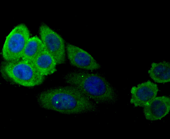
Fig3: ICC staining SERPINC1 in HepG2 cells (green). The nuclear counter stain is DAPI (blue). Cells were fixed in paraformaldehyde, permeabilised with 0.25% Triton X100/PBS.Application
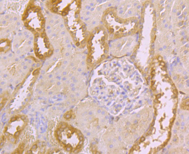
Fig4: Immunohistochemical analysis of paraffin-embedded rat kidney tissue using anti-SERPINC1 antibody. Counter stained with hematoxylin.Application
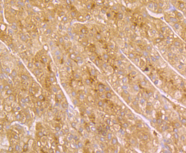
Fig5: Immunohistochemical analysis of paraffin-embedded human liver tissue using anti-SERPINC1 antibody. Counter stained with hematoxylin.Application
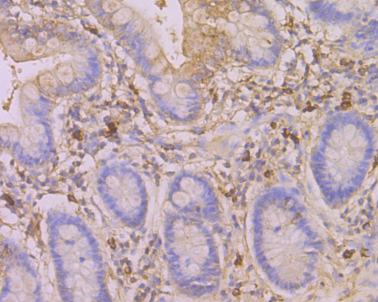
Fig6: Immunohistochemical analysis of paraffin-embedded human small intestine tissue using anti-SERPINC1 antibody. Counter stained with hematoxylin.Application
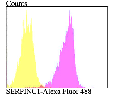
Fig7: Flow cytometric analysis of HepG2 cells with SERPINC1 antibody at 1/100 dilution (purple) compared with an unlabelled control (cells without incubation with primary antibody; yellow). Alexa Fluor 488-conjugated goat anti-rabbit IgG was used as the secondary antibody.| Product Name | SERPINC1 |
|---|---|
| Antibody Type | Primary Antibodies |
| Antigen Alias | ANT3_HUMAN antibody Antithrombin antibody Antithrombin III antibody Antithrombin-III antibody AntithrombinIII antibody AT 3 antibody AT III antibody AT3 antibody AT3D antibody ATIII antibody Heparin cofactor I antibody MGC22579 antibody Serine (or cysteine) proteinase inhibitor clade C (antithrombin) member 1 antibody Serine cysteine proteinase inhibitor clade C member 1 antibody Serine proteinase inhibitor clade C member 1 antibody Serpin C1 antibody Serpin family C member 1 antibody Serpin peptidase inhibitor clade C (antithrombin) member 1 antibody SERPINC1 antibody THPH7 antibody |
| Product description | The serine proteinase inhibitors (serpins) compose a superfamily of proteins with a diverse set of functions, including the control of blood coagulation, complement activation, programmed cell death and development. Serpins are secreted glycoproteins that contain a stretch of peptide that mimics a true substrate for a corresponding serine protease. Antithrombin III (ATIII), an extracellular plasma protein, is a crucial serine protease inhibitor that regulates the coagulation cascade in blood. The inhibitory activity of ATIII is amplified in the presence of heparin. ATIII inhibits Thrombin and Factors IXa, Xa and XIa. Defects in the gene SERPINC1, which encodes for ATIII, can cause ATIII deficiency, an autosomal dominant disease which is a risk factor for hereditary thrombophilia. |
| Immunogen | Recombinant protein within human SERPINC1 aa 150-400. |
| Clonality | Polyclonal |
|---|---|
| Isotype | IgG |
| Host Species | Rabbit |
| Tested Applications | WBICCIHCFC |
| WB:1:500-1 1,000ICC:1:50-1:200 IHC:1:50-1:200 FC:1:50-1:100 |
|
| Species Reactivity | HumanMouseRat |
| Concentration | 1mg/ml |
| Alternative Names | ANT3_HUMAN antibody Antithrombin antibody Antithrombin III antibody Antithrombin-III antibody AntithrombinIII antibody AT 3 antibody AT III antibody AT3 antibody AT3D antibody ATIII antibody Heparin cofactor I antibody MGC22579 antibody Serine (or cysteine) proteinase inhibitor clade C (antithrombin) member 1 antibody Serine cysteine proteinase inhibitor clade C member 1 antibody Serine proteinase inhibitor clade C member 1 antibody Serpin C1 antibody Serpin family C member 1 antibody Serpin peptidase inhibitor clade C (antithrombin) member 1 antibody SERPINC1 antibody THPH7 antibody |
|---|---|
| Molecular Weight(MW) | 53 kDa |
| Cellular Localization | Secreted. |
Application
Fig1: Western blot analysis of SERPINC1 on different lysates using anti-SERPINC1 antibody at 1/500 dilution. Positive control: Lane1: HepG2 Lane2: Mouse smooth muscle Lane3: Mouse lung
Application
Fig2: ICC staining SERPINC1 in H22 cells (green). The nuclear counter stain is DAPI (blue). Cells were fixed in paraformaldehyde, permeabilised with 0.25% Triton X100/PBS.
Application
Fig3: ICC staining SERPINC1 in HepG2 cells (green). The nuclear counter stain is DAPI (blue). Cells were fixed in paraformaldehyde, permeabilised with 0.25% Triton X100/PBS.
Application
Fig4: Immunohistochemical analysis of paraffin-embedded rat kidney tissue using anti-SERPINC1 antibody. Counter stained with hematoxylin.
Application
Fig5: Immunohistochemical analysis of paraffin-embedded human liver tissue using anti-SERPINC1 antibody. Counter stained with hematoxylin.
Application
Fig6: Immunohistochemical analysis of paraffin-embedded human small intestine tissue using anti-SERPINC1 antibody. Counter stained with hematoxylin.
Application
Fig7: Flow cytometric analysis of HepG2 cells with SERPINC1 antibody at 1/100 dilution (purple) compared with an unlabelled control (cells without incubation with primary antibody; yellow). Alexa Fluor 488-conjugated goat anti-rabbit IgG was used as the secondary antibody.| Positive Control | HepG2, mouse smooth muscle tissue, mouse lung tissue, H22, rat kidney tissue, human liver tissue, human small intestine tissue. |
|---|---|
| Application Notes | WB:1:500-1 1,000ICC:1:50-1:200 IHC:1:50-1:200 FC:1:50-1:100 |
| Form | Liquid |
|---|---|
| Storage Instructions | Store at +4℃ after thawing. Aliquot store at -20℃. Avoid repeated freeze / thaw cycles. |
| Storage Buffer | 1*TBS (pH7.4), 0.5%BSA, 50%Glycerol. Preservative: 0.05% Sodium Azide. |


