Application
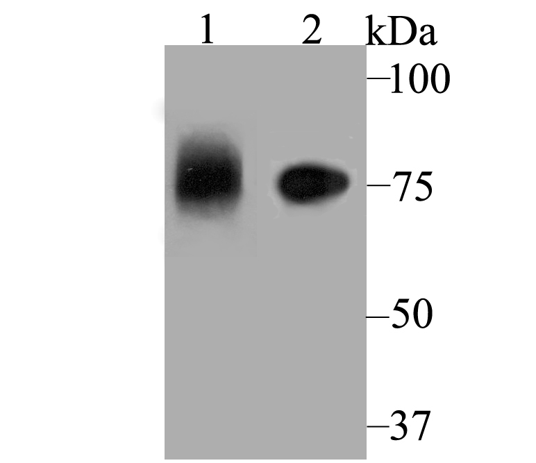
Fig1: Western blot analysis of ApoER2 on different lysates using anti-ApoER2 antibody at 1/1,000 dilution. Positive control: Lane1: Mouse placenta Lane2: SH-SY-5YApplication
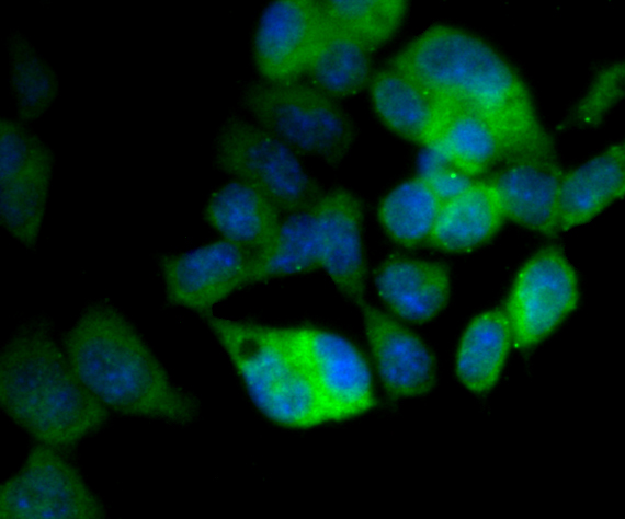
Fig2: ICC staining ApoER2 in F9 cells (green). The nuclear counter stain is DAPI (blue). Cells were fixed in paraformaldehyde, permeabilised with 0.25% Triton X100/PBS.Application
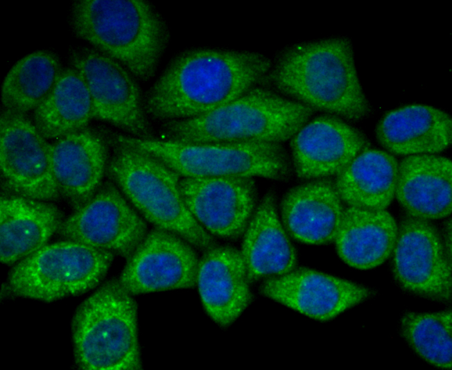
Fig3: ICC staining ApoER2 in HepG2 cells (green). The nuclear counter stain is DAPI (blue). Cells were fixed in paraformaldehyde, permeabilised with 0.25% Triton X100/PBS.Application
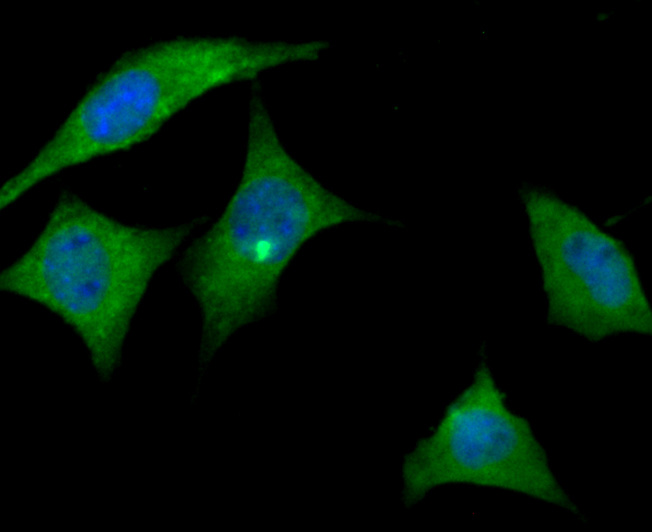
Fig4: ICC staining ApoER2 in SH-SY-5Y cells (green). The nuclear counter stain is DAPI (blue). Cells were fixed in paraformaldehyde, permeabilised with 0.25% Triton X100/PBS.Application
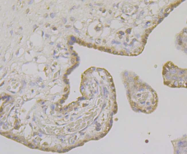
Fig5: Immunohistochemical analysis of paraffin-embedded human placenta tissue using anti-ApoER2 antibody. Counter stained with hematoxylin.Application
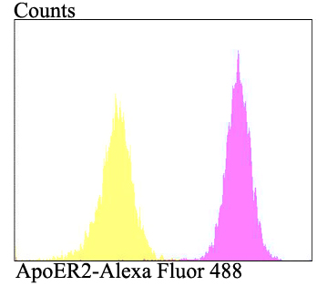
Fig6: Flow cytometric analysis of SH-SY-5Y cells with ApoER2 antibody at 1/100 dilution (purple) compared with an unlabelled control (cells without incubation with primary antibody; yellow). Alexa Fluor 488-conjugated goat anti-rabbit IgG was used as the secondary antibody.| Product Name | ApoER2 |
|---|---|
| Antibody Type | Primary Antibodies |
| Antigen Alias | APOER2 antibody Apolipoprotein E receptor 2 antibody low density lipoprotein receptor-related protein 8 antibody Low-density lipoprotein receptor-related protein 8 antibody LRP-8 antibody LRP8 antibody LRP8_HUMAN antibody |
| Product description | ApoER2 (apolipoprotein E receptor 2), also designated LRP8, is a member of the LDL receptor gene family, which includes LDL receptor, LRP, megalin, VLDLR and ApoER2. The LDL receptor family is characterized by a cluster of cysteine-rich class A repeats, epidermal growth factor (EGF)-like repeats, YWTD repeats and an O-linked sugar domain. ApoER2 is expressed in brain and placenta and has several splice variants. ApoER2 is thought to mediate the interaction of extracellular Reelin and cytosolic mDab1 (mammalian disabled protein), which activates a tyrosine kinase. This pathway regulates the migration of neurons along the radial glial fiber network during brain development. |
| Immunogen | Recombinant protein within human ApoER2 500-750 aa. |
| Clonality | Polyclonal |
|---|---|
| Isotype | IgG |
| Host Species | Rabbit |
| Tested Applications | WBICCIHCFC |
| WB:1:500-1:1,000 ICC:1:50-1:200 IHC:1:50 FC:1:50-1:100 |
|
| Species Reactivity | HumanMouse |
| Concentration | 1mg/ml |
| Alternative Names | APOER2 antibody Apolipoprotein E receptor 2 antibody low density lipoprotein receptor-related protein 8 antibody Low-density lipoprotein receptor-related protein 8 antibody LRP-8 antibody LRP8 antibody LRP8_HUMAN antibody |
|---|---|
| Molecular Weight(MW) | 77 kDa |
| Cellular Localization | Cell membrane. Secreted. |
Application
Fig1: Western blot analysis of ApoER2 on different lysates using anti-ApoER2 antibody at 1/1,000 dilution. Positive control: Lane1: Mouse placenta Lane2: SH-SY-5Y
Application
Fig2: ICC staining ApoER2 in F9 cells (green). The nuclear counter stain is DAPI (blue). Cells were fixed in paraformaldehyde, permeabilised with 0.25% Triton X100/PBS.
Application
Fig3: ICC staining ApoER2 in HepG2 cells (green). The nuclear counter stain is DAPI (blue). Cells were fixed in paraformaldehyde, permeabilised with 0.25% Triton X100/PBS.
Application
Fig4: ICC staining ApoER2 in SH-SY-5Y cells (green). The nuclear counter stain is DAPI (blue). Cells were fixed in paraformaldehyde, permeabilised with 0.25% Triton X100/PBS.
Application
Fig5: Immunohistochemical analysis of paraffin-embedded human placenta tissue using anti-ApoER2 antibody. Counter stained with hematoxylin.
Application
Fig6: Flow cytometric analysis of SH-SY-5Y cells with ApoER2 antibody at 1/100 dilution (purple) compared with an unlabelled control (cells without incubation with primary antibody; yellow). Alexa Fluor 488-conjugated goat anti-rabbit IgG was used as the secondary antibody.| Positive Control | Mouse placenat tissue, SH-SY-5Y, F9, HepG2, human placenta tissue. |
|---|---|
| Application Notes | WB:1:500-1:1,000 ICC:1:50-1:200 IHC:1:50 FC:1:50-1:100 |
| Form | Liquid |
|---|---|
| Storage Instructions | Store at +4℃ after thawing. Aliquot store at -20℃. Avoid repeated freeze / thaw cycles. |
| Storage Buffer | 1*TBS (pH7.4), 0.5%BSA, 50%Glycerol. Preservative: 0.05% Sodium Azide. |


