WB
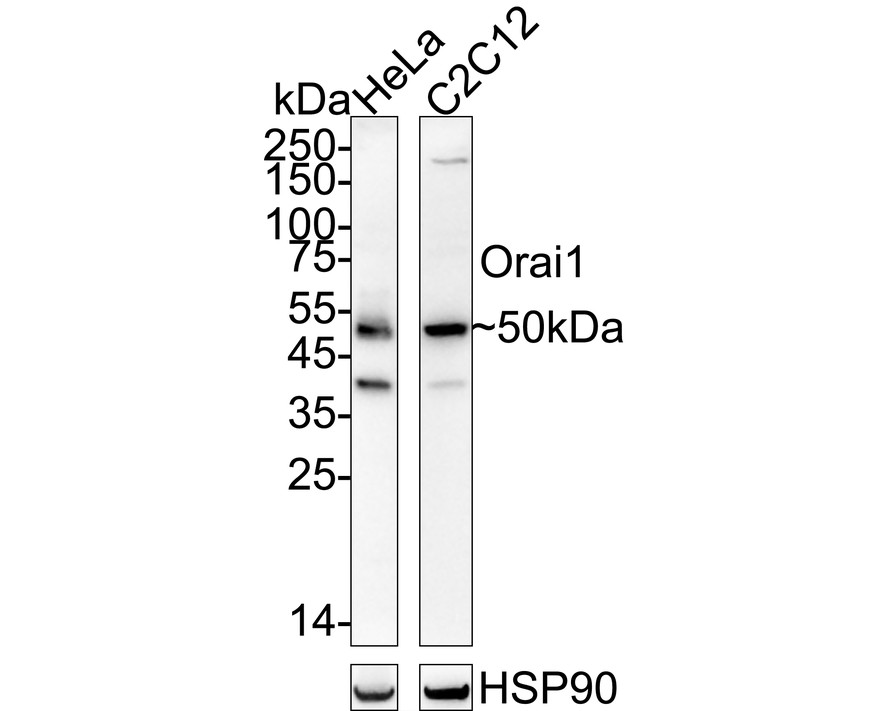
Western blot analysis of Orai1 on different lysates with Rabbit anti-Orai1 antibody at 1/1,000 dilution. Lane 1: HeLa cell lysate, Lane 2: C2C12 cell lysate, Lysates/proteins at 20 µg/Lane. Exposure time: 59 seconds; ECL: K1801; 4-20% SDS-PAGE gel. Proteins were transferred to a PVDF membrane and blocked with 5% NFDM/TBST for 1 hour at room temperature. The primary antibody at 1/1,000 dilution was used in 5% NFDM/TBST at 4℃ overnight. Goat Anti-Rabbit IgG - HRP Secondary Antibody at 1/50,000 dilution was used for 1 hour at room temperature.IHC
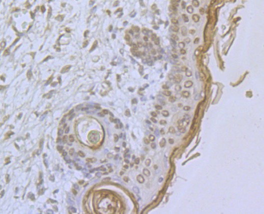
Immunohistochemical analysis of paraffin-embedded human skin tissue using anti- Orai1 antibody. Counter stained with hematoxylin.IHC
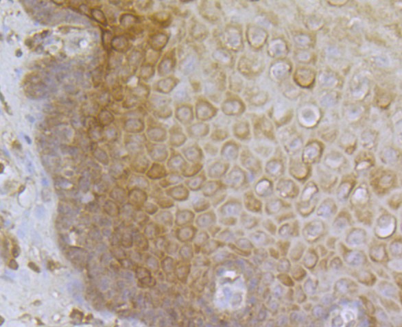
Immunohistochemical analysis of paraffin-embedded human esophagus tissue using anti-Orai1 antibody. Counter stained with hematoxylin.| Product Name | Orai1 Rabbit Polyclonal Antibody |
|---|---|
| Antibody Type | Primary Antibodies |
| Product description | Orai1 (ORAI calcium release-activated calcium modulator 1), also known as ORAT1, CRACM1 (calcium release-activated calcium modulator 1) or TMEM142A (transmembrane protein 142A), is a 301 amino acid multi-pass membrane protein that belongs to the Orai family of proteins. Localizing to the plasma membrane, Orai1 plays an important role in store-operated calcium (SOC) entry, a process involving Ca2+ influx and replenishment of Ca2+ stores formerly emptied through the action of inositol 1,4,5-trisphosphate production and other Ca2+ mobilizing agents. Specifically, Orai1 functions as a pore subunit of the store-operated calcium release-activated calcium channel (CRAC) and is essential for proper function of the CRAC channel. CRAC channels are responsible for mediating calcium influx in T-cells and play an important role in the immune response. Mutations in the gene encoding Orai1 can result in severe combined immunodeficiency (SCID). |
| Immunogen | Synthetic peptide within mouse Orai1 aa 150-250. |
| Clonality | Polyclonal |
|---|---|
| Isotype | IgG |
| Host Species | Rabbit |
| Tested Applications | ICC/IFIHCWB |
| WB:1:500-1:2,000 ICC:1:50-1:200 IHC:1:50-1:200 |
|
| Species Reactivity | HumanMouse |
| Concentration | 1mg/ml |
| Purification | Affinity purified |
| Alternative Names | Calcium release activated calcium channel protein 1 antibody Calcium release activated calcium modulator 1 antibody Calcium release-activated calcium channel protein 1 antibody CRACM 1 antibody CRACM1 antibody CRCM1 antibody CRCM1_HUMAN antibody FLJ14466 antibody Orai 1 antibody ORAI calcium release activated calcium modulator 1 antibody orai1 antibody ORAT1 antibody Protein orai 1 antibody Protein orai-1 antibody TMEM 142A antibody TMEM142A antibody Transmembrane protein 142A antibody |
|---|---|
| Molecular Weight(MW) | 33 kDa(Observed band size: 50 kDa) |
| Cellular Localization | Cell membrane. |
WB
Western blot analysis of Orai1 on different lysates with Rabbit anti-Orai1 antibody at 1/1,000 dilution. Lane 1: HeLa cell lysate, Lane 2: C2C12 cell lysate, Lysates/proteins at 20 µg/Lane. Exposure time: 59 seconds; ECL: K1801; 4-20% SDS-PAGE gel. Proteins were transferred to a PVDF membrane and blocked with 5% NFDM/TBST for 1 hour at room temperature. The primary antibody at 1/1,000 dilution was used in 5% NFDM/TBST at 4℃ overnight. Goat Anti-Rabbit IgG - HRP Secondary Antibody at 1/50,000 dilution was used for 1 hour at room temperature.
IHC
Immunohistochemical analysis of paraffin-embedded human skin tissue using anti- Orai1 antibody. Counter stained with hematoxylin.
IHC
Immunohistochemical analysis of paraffin-embedded human esophagus tissue using anti-Orai1 antibody. Counter stained with hematoxylin.| Application Notes | WB:1:500-1:2,000 ICC:1:50-1:200 IHC:1:50-1:200 |
|---|
| Form | Liquid |
|---|---|
| Storage Instructions | Store at +4℃ after thawing. Aliquot store at -20℃. Avoid repeated freeze / thaw cycles. |
| Storage Buffer | 1*TBS (pH7.4), 0.5%BSA, 50%Glycerol. Preservative: 0.05% Sodium Azide. |


