Application
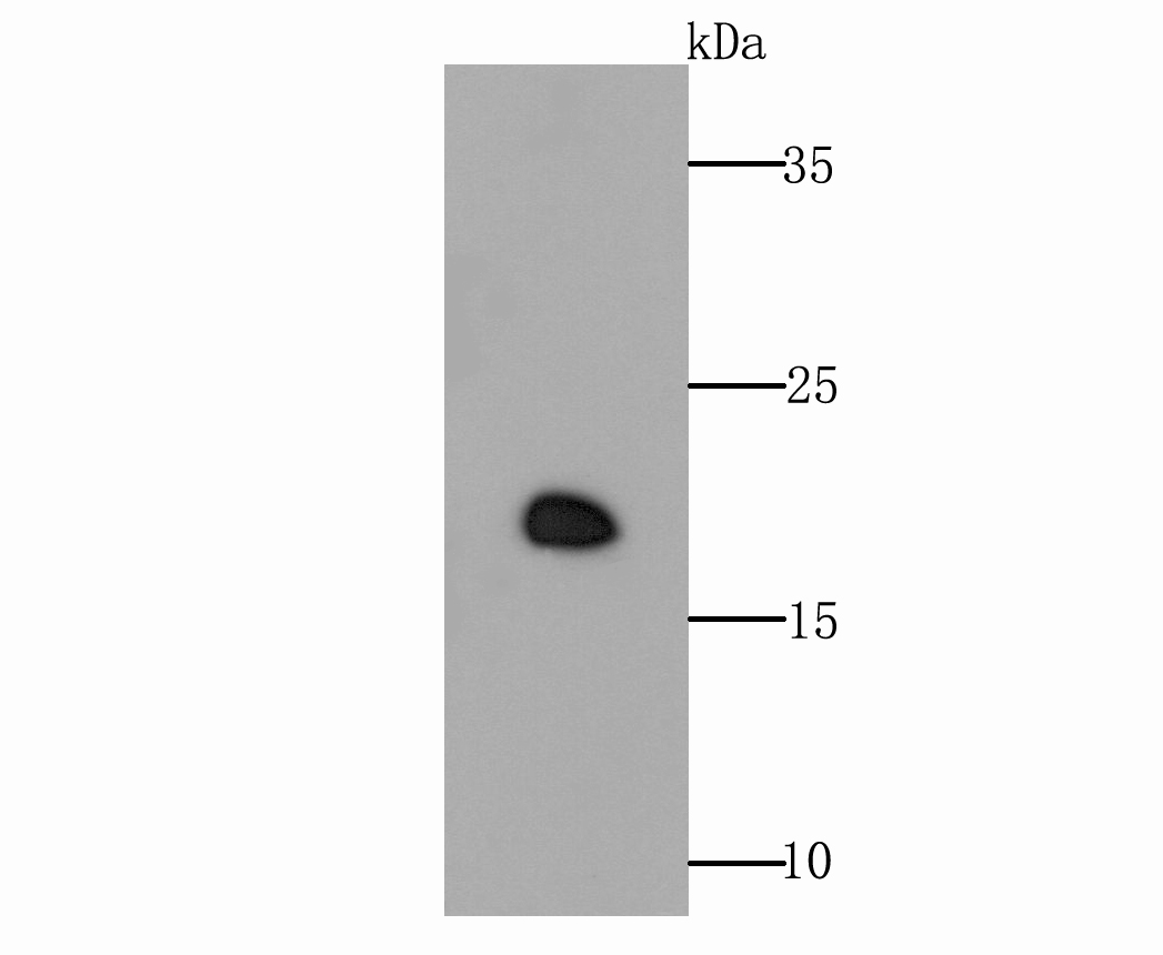
Fig1: Western blot analysis of GPX4 on rat testis tissue lysate using anti- GPX4 antibody at 1/500 dilution.Application
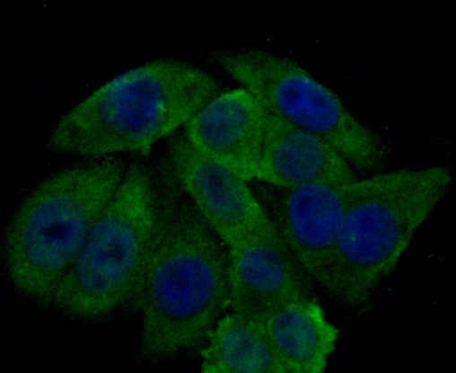
Fig2: ICC staining GPX4 in HepG2 cells (green). The nuclear counter stain is DAPI (blue). Cells were fixed in paraformaldehyde, permeabilised with 0.25% Triton X100/PBS.Application
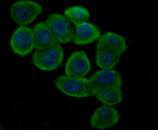
Fig3: ICC staining GPX4 in LOVO cells (green). The nuclear counter stain is DAPI (blue). Cells were fixed in paraformaldehyde, permeabilised with 0.25% Triton X100/PBS.Application
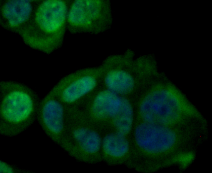
Fig4: ICC staining GPX4 in MCF-7 cells (green). The nuclear counter stain is DAPI (blue). Cells were fixed in paraformaldehyde, permeabilised with 0.25% Triton X100/PBS.Application
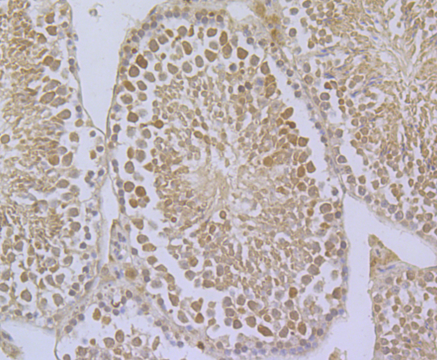
Fig5: Immunohistochemical analysis of paraffin-embedded mouse testis tissue using anti- GPX4 antibody. Counter stained with hematoxylin.Application
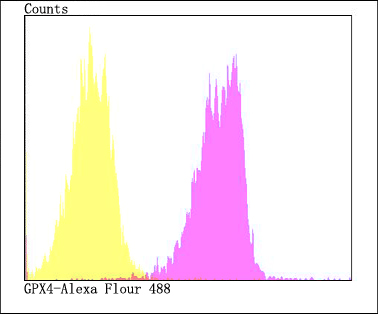
Fig6: Flow cytometric analysis of HepG2 cells with GPX4 antibody at 1/100 dilution (fuchsia) compared with an unlabelled control (cells without incubation with primary antibody; yellow). Alexa Fluor 488-conjugated goat anti-rabbit IgG was used as the secondary antibody.| Product Name | GPX4 |
|---|---|
| Antibody Type | Primary Antibodies |
| Antigen Alias | Glutathione peroxidase 4 antibody GPX 4 antibody GPX-4 antibody GPX4 antibody GPX4_HUMAN antibody GSHPx-4 antibody MCSP antibody mitochondrial antibody PHGPx antibody Phospholipid hydroperoxidase antibody Phospholipid hydroperoxide glutathione peroxidase antibody Phospholipid hydroperoxide glutathione peroxidase mitochondrial antibody snGPx antibody snPHGPx antibody Sperm nucleus glutathione peroxidase antibody |
| Product description | Essential antioxidant peroxidase that directly reduces phospholipid hydroperoxide even if they are incorporated in membranes and lipoproteins . Can also reduce fatty acid hydroperoxide, cholesterol hydroperoxide and thymine hydroperoxide . Plays a key role in protecting cells from oxidative damage by preventing membrane lipid peroxidation. Required to prevent cells from ferroptosis, a non-apoptotic cell death resulting from an iron-dependent accumulation of lipid reactive oxygen species . The presence of selenocysteine (Sec) versus Cys at the active site is essential for life: it provides resistance to overoxidation and prevents cells against ferroptosis . The presence of Sec at the active site is also essential for the survival of a specific type of parvalbumin-positive interneurons, thereby preventing against fatal epileptic seizures . May be required to protect cells from the toxicity of ingested lipid hydroperoxides . Required for normal sperm development and male fertility. Essential for maturation and survival of photoreceptor cells. Plays a role in a primary T-cell response to viral and parasitic infection by protecting T-cells from ferroptosis and by supporting T-cell expansion. |
| Immunogen | Recombinant protein within N-terminal human GPX4. |
| Clonality | Polyclonal |
|---|---|
| Isotype | IgG |
| Host Species | Rabbit |
| Tested Applications | WBICCIHCFC |
| WB:1:500-1:1,000 ICC:1:50-1:100 IHC:1:50 -1:200 FC:1:50-1:100 |
|
| Species Reactivity | HumanMouseRat |
| Concentration | 1mg/ml |
| Alternative Names | Glutathione peroxidase 4 antibody GPX 4 antibody GPX-4 antibody GPX4 antibody GPX4_HUMAN antibody GSHPx-4 antibody MCSP antibody mitochondrial antibody PHGPx antibody Phospholipid hydroperoxidase antibody Phospholipid hydroperoxide glutathione peroxidase antibody Phospholipid hydroperoxide glutathione peroxidase mitochondrial antibody snGPx antibody snPHGPx antibody Sperm nucleus glutathione peroxidase antibody |
|---|---|
| Molecular Weight(MW) | 22 kDa |
| Cellular Localization | Cytoplasm. Mitochondrion. |
Application
Fig1: Western blot analysis of GPX4 on rat testis tissue lysate using anti- GPX4 antibody at 1/500 dilution.
Application
Fig2: ICC staining GPX4 in HepG2 cells (green). The nuclear counter stain is DAPI (blue). Cells were fixed in paraformaldehyde, permeabilised with 0.25% Triton X100/PBS.
Application
Fig3: ICC staining GPX4 in LOVO cells (green). The nuclear counter stain is DAPI (blue). Cells were fixed in paraformaldehyde, permeabilised with 0.25% Triton X100/PBS.
Application
Fig4: ICC staining GPX4 in MCF-7 cells (green). The nuclear counter stain is DAPI (blue). Cells were fixed in paraformaldehyde, permeabilised with 0.25% Triton X100/PBS.
Application
Fig5: Immunohistochemical analysis of paraffin-embedded mouse testis tissue using anti- GPX4 antibody. Counter stained with hematoxylin.
Application
Fig6: Flow cytometric analysis of HepG2 cells with GPX4 antibody at 1/100 dilution (fuchsia) compared with an unlabelled control (cells without incubation with primary antibody; yellow). Alexa Fluor 488-conjugated goat anti-rabbit IgG was used as the secondary antibody.| Positive Control | Rat testis tissue lysate, HepG2, LOVO, MCF-7, mouse testis tissue. |
|---|---|
| Application Notes | WB:1:500-1:1,000 ICC:1:50-1:100 IHC:1:50 -1:200 FC:1:50-1:100 |
| Form | Liquid |
|---|---|
| Storage Instructions | Store at +4℃ after thawing. Aliquot store at -20℃. Avoid repeated freeze / thaw cycles. |
| Storage Buffer | 1*TBS (pH7.4), 0.5%BSA, 50%Glycerol. Preservative: 0.05% Sodium Azide. |


