Application
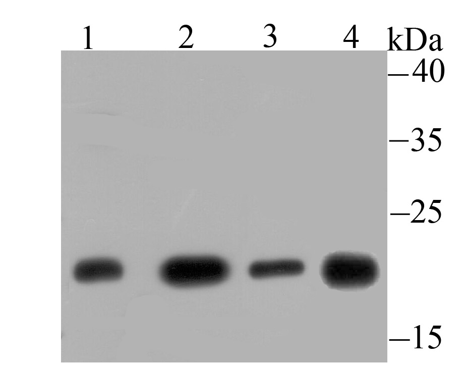
Fig1: Western blot analysis of GLO1 on different lysates using anti-GLO1 antibody at 1/500 dilution. Positive control: Lane 1: Mouse testis tissue Lane 2: C2C12 Lane 3: 293 Lane 4: Rat spleen tissueApplication
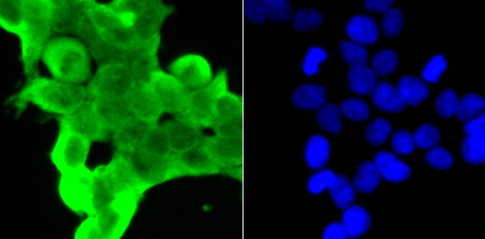
Fig2: ICC staining GLO1 in 293T cells (green). The nuclear counter stain is DAPI (blue). Cells were fixed in paraformaldehyde, permeabilised with 0.25% Triton X100/PBS.Application
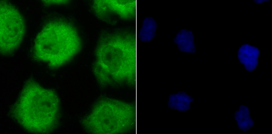
Fig3: ICC staining GLO1 in HUVEC cells (green). The nuclear counter stain is DAPI (blue). Cells were fixed in paraformaldehyde, permeabilised with 0.25% Triton X100/PBS.Application
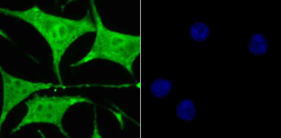
Fig4: ICC staining GLO1 in SH-SY5Y cells (green). The nuclear counter stain is DAPI (blue). Cells were fixed in paraformaldehyde, permeabilised with 0.25% Triton X100/PBS.Application
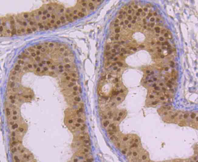
Fig5: Immunohistochemical analysis of paraffin-embedded rat epididymis tissue using anti-GLO1 antibody. Counter stained with hematoxylin.Application
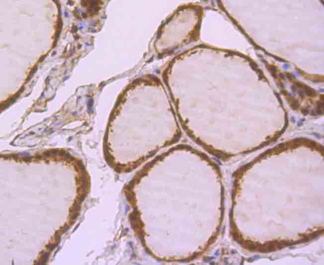
Fig6: Immunohistochemical analysis of paraffin-embedded human thyroid gland tissue using anti-GLO1 antibody. Counter stained with hematoxylin.Application
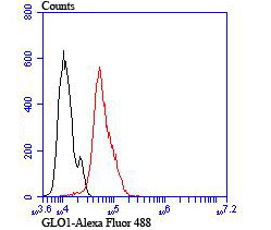
Fig7: Flow cytometric analysis of Hela cells with GLO1 antibody at 1/100 dilution (red) compared with an unlabelled control (cells without incubation with primary antibody; black).| Product Name | GLO1 [JU44-11] |
|---|---|
| Antibody Type | Primary Antibodies |
| Antigen Alias | Aldoketomutase antibody glo1 antibody GLOD1 antibody Glx I antibody GLYI antibody glyoxalase domain containing 1 antibody Glyoxalase I antibody Ketone aldehyde mutase antibody Ketone-aldehyde mutase antibody Lactoyl glutathione lyase antibody Lactoylglutathione lyase antibody LGUL_HUMAN antibody Methylglyoxalase antibody S D lactoylglutathione methylglyoxal lyase antibody S-D-lactoylglutathione methylglyoxal lyase antibody |
| Product description | The glyoxal pathway plays a role in the detoxification of glucose degradation products (GDP). Glyoxalase I (GLO1), a member of the glyoxalase family, is effective in eliminating GDP. Overexpression or silencing of Glyoxalase I in mice brain suggests an association between Glyoxalase I and anxiety. Glyoxalase I has three isoforms generated from two alleles in the genome which forms two homodimers and one heterodimer, each subunit binding one zinc ion. Research demonstrates that GLO1 gene expression is induced in colon carcinoma. Both an insulin response element (IRE), and a zinc metal response element (MRE) in the promoter region of the GLO1 gene have been identified. |
| Immunogen | Recombinant protein |
| Clonality | Monoclonal |
|---|---|
| Isotype | IgG |
| Host Species | Recombinant rabbit |
| Tested Applications | WBICCIHCFC |
| WB:1:500-1:2,000 ICC:1:100-1:500 IHC:1:100-1:500 FC:1:50-1:100 |
|
| Species Reactivity | HumanMouseRat |
| Concentration | 1mg/ml |
| Alternative Names | Aldoketomutase antibody glo1 antibody GLOD1 antibody Glx I antibody GLYI antibody glyoxalase domain containing 1 antibody Glyoxalase I antibody Ketone aldehyde mutase antibody Ketone-aldehyde mutase antibody Lactoyl glutathione lyase antibody Lactoylglutathione lyase antibody LGUL_HUMAN antibody Methylglyoxalase antibody S D lactoylglutathione methylglyoxal lyase antibody S-D-lactoylglutathione methylglyoxal lyase antibody |
|---|---|
| Molecular Weight(MW) | 21 kDa |
| Cellular Localization | Cytoplasm. Nucleus. |
Application
Fig1: Western blot analysis of GLO1 on different lysates using anti-GLO1 antibody at 1/500 dilution. Positive control: Lane 1: Mouse testis tissue Lane 2: C2C12 Lane 3: 293 Lane 4: Rat spleen tissue
Application
Fig2: ICC staining GLO1 in 293T cells (green). The nuclear counter stain is DAPI (blue). Cells were fixed in paraformaldehyde, permeabilised with 0.25% Triton X100/PBS.
Application
Fig3: ICC staining GLO1 in HUVEC cells (green). The nuclear counter stain is DAPI (blue). Cells were fixed in paraformaldehyde, permeabilised with 0.25% Triton X100/PBS.
Application
Fig4: ICC staining GLO1 in SH-SY5Y cells (green). The nuclear counter stain is DAPI (blue). Cells were fixed in paraformaldehyde, permeabilised with 0.25% Triton X100/PBS.
Application
Fig5: Immunohistochemical analysis of paraffin-embedded rat epididymis tissue using anti-GLO1 antibody. Counter stained with hematoxylin.
Application
Fig6: Immunohistochemical analysis of paraffin-embedded human thyroid gland tissue using anti-GLO1 antibody. Counter stained with hematoxylin.
Application
Fig7: Flow cytometric analysis of Hela cells with GLO1 antibody at 1/100 dilution (red) compared with an unlabelled control (cells without incubation with primary antibody; black).| Positive Control | Mouse testis tissue lysate, C2C12, 293, rat spleen tissue lysate, 293T, HUVEC, SH-SY5Y, Hela, rat epididymis tissue, human thyroid gland tissue. |
|---|---|
| Application Notes | WB:1:500-1:2,000 ICC:1:100-1:500 IHC:1:100-1:500 FC:1:50-1:100 |
| Form | Liquid |
|---|---|
| Storage Instructions | Store at +4℃ after thawing. Aliquot store at -20℃ or -80℃. Avoid repeated freeze / thaw cycles. |
| Storage Buffer | 1*TBS (pH7.4), 1%BSA, 40%Glycerol. Preservative: 0.05% Sodium Azide. |


