Application
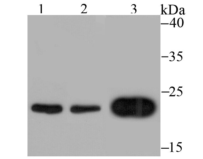
Fig1: Western blot analysis of Mad2L1 on different cell lysates using anti-Mad2L1 antibody at 1/500 dilution. Positive control: Lane 1: K562 Lane 2: 293T Lane 3: SH-SY5YApplication
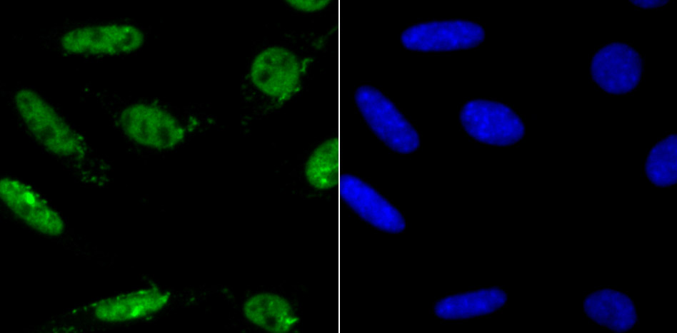
Fig2: ICC staining Mad2L1 in SH-SY5Y cells (green). The nuclear counter stain is DAPI (blue). Cells were fixed in paraformaldehyde, permeabilised with 0.25% Triton X100/PBS.| Product Name | Mad2L1 [JU46-13] |
|---|---|
| Antibody Type | Primary Antibodies |
| Antigen Alias | Caught by MAD2 protein antibody CMT2 antibody dJ261G23.1 antibody KIAA0110 antibody MAD2L1 binding protein antibody MAD2L1 binding protein isoform 2 antibody MAD2L1-binding protein antibody Mad2l1bp antibody MD2BP_HUMAN antibody MGC11282 antibody OTTHUMP00000016496 antibody RP1 261G23.6 antibody |
| Product description | Cell cycle progression is subject to arrest at the mitotic spindle assembly checkpoint in response to incorrect spindle fiber assembly. MAD2 (for mitotic arrest-deficient) is a component of the mitotic spindle checkpoint. Cells with mutated MAD2 do not undergo mitotic arrest in response to incorrect spindle fiber assembly, which results in missegregation and eventual cell death. A breast carcinoma cell line with reduced MAD2 expression, T47D, was shown to complete mitosis in the presence of nocodazole, an inhibitor of mitotic spindle assembly. MAD2 is localized to unattached kinetochores during prometaphase and disassociates upon spindle fiber attachment, indicating that MAD2 regulates kinetochore binding to the spindle fibers. Human MAD2 has also been shown to associate with insulin receptor (IR), but not IGFIR, implicating MAD2 as a mediator for IR-specific signaling. MAD2B, a MAD2 homolog, is required for the execution of the mitotic checkpoint monitoring the kinetochore-spindle attachment process and if the process is not complete, MAD2B delays the onset of anaphase. |
| Immunogen | Recombinant protein |
| Clonality | Monoclonal |
|---|---|
| Isotype | IgG |
| Host Species | Recombinant rabbit |
| Tested Applications | WBICC |
| WB:1:500-1:2,000 ICC:1:100-1:500 |
|
| Species Reactivity | Human |
| Concentration | 1mg/ml |
| Alternative Names | Caught by MAD2 protein antibody CMT2 antibody dJ261G23.1 antibody KIAA0110 antibody MAD2L1 binding protein antibody MAD2L1 binding protein isoform 2 antibody MAD2L1-binding protein antibody Mad2l1bp antibody MD2BP_HUMAN antibody MGC11282 antibody OTTHUMP00000016496 antibody RP1 261G23.6 antibody |
|---|---|
| Molecular Weight(MW) | 23.5 kDa |
| Cellular Localization | Nucleus. Cytoskeleton. |
| SwissProt ID | Q13257 |
|---|
Application
Fig1: Western blot analysis of Mad2L1 on different cell lysates using anti-Mad2L1 antibody at 1/500 dilution. Positive control: Lane 1: K562 Lane 2: 293T Lane 3: SH-SY5Y
Application
Fig2: ICC staining Mad2L1 in SH-SY5Y cells (green). The nuclear counter stain is DAPI (blue). Cells were fixed in paraformaldehyde, permeabilised with 0.25% Triton X100/PBS.| Positive Control | K562, 293T, SH-SY5Y. |
|---|---|
| Application Notes | WB:1:500-1:2,000 ICC:1:100-1:500 |
| Form | Liquid |
|---|---|
| Storage Instructions | Store at +4℃ after thawing. Aliquot store at -20℃ or -80℃. Avoid repeated freeze / thaw cycles. |
| Storage Buffer | 1*TBS (pH7.4), 1%BSA, 40%Glycerol. Preservative: 0.05% Sodium Azide. |


