Application
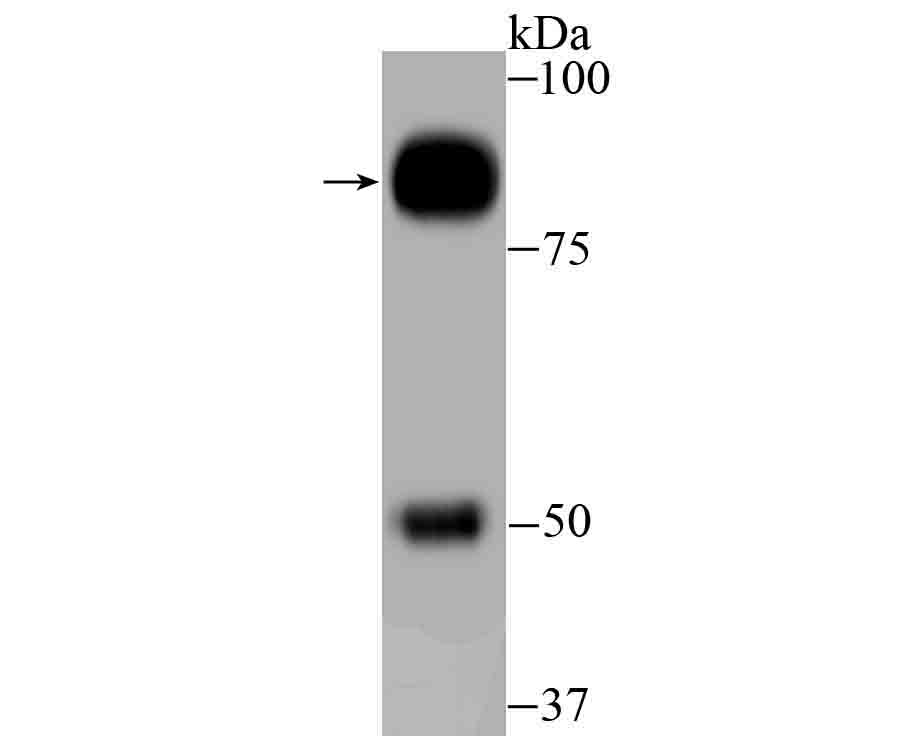
Fig1: Western blot analysis of GAB1 on human small intestine tissue lysate using anti-GAB1 antibody at 1/500 dilution.Application
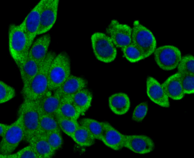
Fig2: ICC staining GAB1 in LOVO cells (green). The nuclear counter stain is DAPI (blue). Cells were fixed in paraformaldehyde, permeabilised with 0.25% Triton X100/PBS.Application
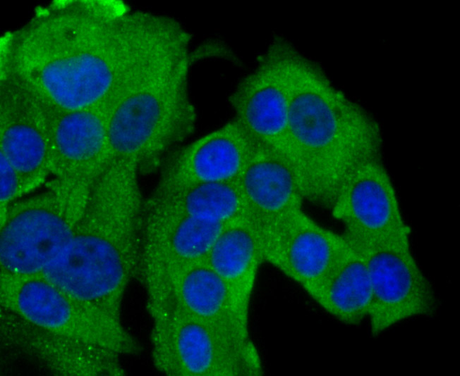
Fig3: ICC staining GAB1 in MCF-7 cells (green). The nuclear counter stain is DAPI (blue). Cells were fixed in paraformaldehyde, permeabilised with 0.25% Triton X100/PBS.Application
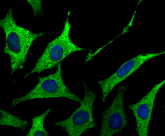
Fig4: ICC staining GAB1 in SH-SY5Y cells (green). The nuclear counter stain is DAPI (blue). Cells were fixed in paraformaldehyde, permeabilised with 0.25% Triton X100/PBS.Application
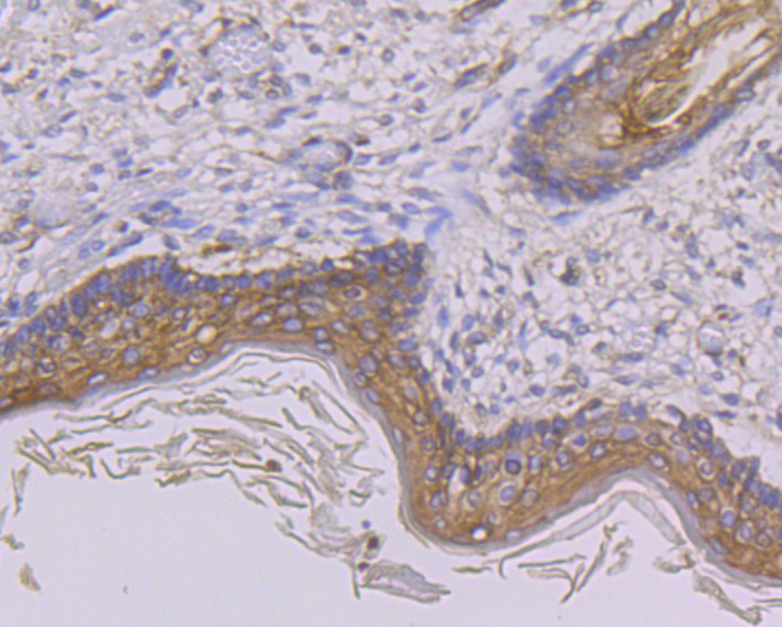
Fig5: Immunohistochemical analysis of paraffin-embedded human skin tissue using anti-GAB1 antibody. Counter stained with hematoxylin.Application
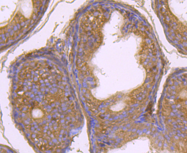
Fig6: Immunohistochemical analysis of paraffin-embedded rat epididymis tissue using anti-GAB1 antibody. Counter stained with hematoxylin.Application
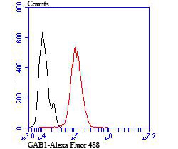
Fig7: Flow cytometric analysis of Hela cells with GAB1 antibody at 1/100 dilution (red) compared with an unlabelled control (cells without incubation with primary antibody; black). Alexa Fluor 488-conjugated goat anti-rabbit IgG was used as the secondary antibody.| Product Name | GAB1 [JU47-19] |
|---|---|
| Antibody Type | Primary Antibodies |
| Antigen Alias | Gab 1 antibody GAB1 antibody GAB1_HUMAN antibody GRB 2 associated binder 1 antibody GRB 2 associated binding protein 1 antibody GRB2 associated binding protein 1 isoform a antibody GRB2 associated binding protein 1 isoform b antibody GRB2-associated binder 1 antibody GRB2-associated-binding protein 1 antibody Growth factor receptor bound protein 2-associated protein 1 antibody |
| Product description | Growth factor triggering of protein tyrosine kinase receptors induces signals that cascade to the nucleus, activating mitogenic as well as other responses. Critical components of this process include adapter proteins such as Shc, IRS-1 and Gab 1 (GRB-associated binder-1) that lack detectable catalytic activity. These are immediate substrates of receptor tyrosine kinase activity and serve to link activated receptors to downstream signaling components. Whereas Shc has been implicated in signaling by diverse receptor families, IRS-1 serves primarily as the major insulin receptor substrate. Shc and Gab 1 also participate in insulin signaling by linking the insulin receptor to Ras by forming complexes with GRB2 (another adapter protein) and Sos independently of IRS-1. Gab 1 is also thought to be involved in the EGF receptor signaling pathway. |
| Immunogen | Recombinant protein |
| Clonality | Monoclonal |
|---|---|
| Isotype | IgG |
| Host Species | Recombinant rabbit |
| Tested Applications | WBICCIFIHCFC |
| WB:1:500-1:2,000 ICC:1:100-1:500 IHC:1:50-1:200 FC:1:50-1:100 |
|
| Species Reactivity | HumanMouseRat |
| Concentration | 1mg/ml |
| Alternative Names | Gab 1 antibody GAB1 antibody GAB1_HUMAN antibody GRB 2 associated binder 1 antibody GRB 2 associated binding protein 1 antibody GRB2 associated binding protein 1 isoform a antibody GRB2 associated binding protein 1 isoform b antibody GRB2-associated binder 1 antibody GRB2-associated-binding protein 1 antibody Growth factor receptor bound protein 2-associated protein 1 antibody |
|---|---|
| Molecular Weight(MW) | 77 kDa |
| Cellular Localization | Cytosol. |
| SwissProt ID | Q13480 |
|---|
Application
Fig1: Western blot analysis of GAB1 on human small intestine tissue lysate using anti-GAB1 antibody at 1/500 dilution.
Application
Fig2: ICC staining GAB1 in LOVO cells (green). The nuclear counter stain is DAPI (blue). Cells were fixed in paraformaldehyde, permeabilised with 0.25% Triton X100/PBS.
Application
Fig3: ICC staining GAB1 in MCF-7 cells (green). The nuclear counter stain is DAPI (blue). Cells were fixed in paraformaldehyde, permeabilised with 0.25% Triton X100/PBS.
Application
Fig4: ICC staining GAB1 in SH-SY5Y cells (green). The nuclear counter stain is DAPI (blue). Cells were fixed in paraformaldehyde, permeabilised with 0.25% Triton X100/PBS.
Application
Fig5: Immunohistochemical analysis of paraffin-embedded human skin tissue using anti-GAB1 antibody. Counter stained with hematoxylin.
Application
Fig6: Immunohistochemical analysis of paraffin-embedded rat epididymis tissue using anti-GAB1 antibody. Counter stained with hematoxylin.
Application
Fig7: Flow cytometric analysis of Hela cells with GAB1 antibody at 1/100 dilution (red) compared with an unlabelled control (cells without incubation with primary antibody; black). Alexa Fluor 488-conjugated goat anti-rabbit IgG was used as the secondary antibody.| Positive Control | Human small intestine tissue lysate, LOVO, MCF-7, SH-SY5Y, human skin tissue, rat epididymis tissue, Hela. |
|---|---|
| Application Notes | WB:1:500-1:2,000 ICC:1:100-1:500 IHC:1:50-1:200 FC:1:50-1:100 |
| Form | Liquid |
|---|---|
| Storage Instructions | Store at +4℃ after thawing. Aliquot store at -20℃ or -80℃. Avoid repeated freeze / thaw cycles. |
| Storage Buffer | 1*TBS (pH7.4), 1%BSA, 40%Glycerol. Preservative: 0.05% Sodium Azide. |


