Application
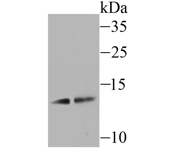
Fig1: Western blot analysis of SCGB1A1 on mouse lung tissue (1) and A549 cell (2) lysate using anti-SCGB1A1 antibody at 1/500 dilution.Application
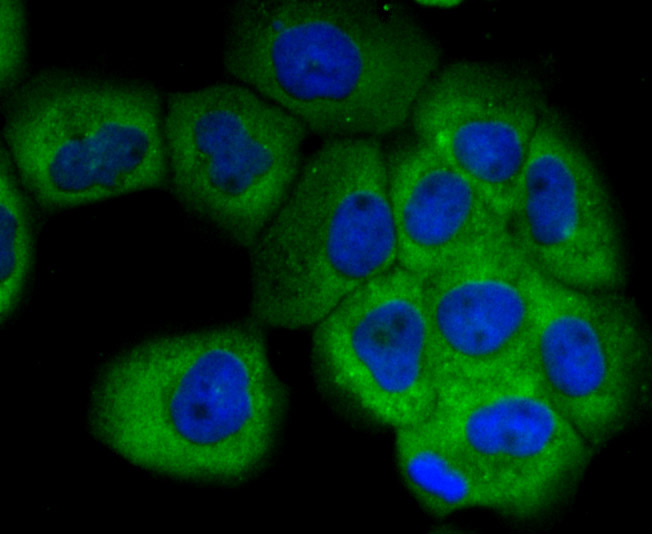
Fig2: ICC staining SCGB1A1 in A431 cells (green). The nuclear counter stain is DAPI (blue). Cells were fixed in paraformaldehyde, permeabilised with 0.25% Triton X100/PBS.Application
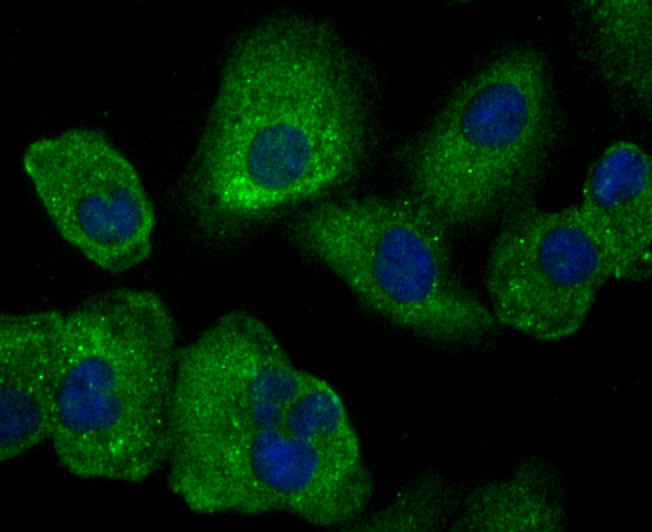
Fig3: ICC staining SCGB1A1 in A549 cells (green). The nuclear counter stain is DAPI (blue). Cells were fixed in paraformaldehyde, permeabilised with 0.25% Triton X100/PBS.Application
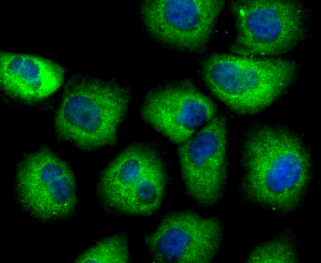
Fig4: ICC staining SCGB1A1 in HUVEC cells (green). The nuclear counter stain is DAPI (blue). Cells were fixed in paraformaldehyde, permeabilised with 0.25% Triton X100/PBS.Application
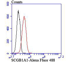
Fig5: Flow cytometric analysis of A549 cells with SCGB1A1 antibody at 1/100 dilution (red) compared with an unlabelled control (cells without incubation with primary antibody; black).| Product Name | SCGB1A1 [JU34-03] |
|---|---|
| Antibody Type | Primary Antibodies |
| Antigen Alias | Blastokinin antibody CC10 antibody CC16 antibody CCPBP antibody CCSP antibody Clara cell phospholipid binding protein antibody Clara cell phospholipid-binding protein antibody Clara cell specific 10 kD protein antibody Clara cells 10 kDa secretory protein antibody OTTHUMP00000236107 antibody SCGB1A1 antibody Secretoglobin family 1A member 1 antibody Secretoglobin, family 1A, member 1 (uteroglobin) antibody UG antibody UGB antibody UP-1 antibody UP1 antibody Urinary protein 1 antibody Urine protein 1 antibody UTER_HUMAN antibody Uteroglobin antibody |
| Product description | SCGB1A1 also named CC10. Clara cell 10 (CC10) protein, a homologue of rabbit uteroglobin, is a phospholipase A2 inhibitor. CC10 is regulated by AP-1, octamer, and hepatocyte nuclear factor-3 (HNF-3) family transcription factors. CC10 expression changes in relation to the ovarian menstrual cycle, and expression in human endometrium may be stimulated by progesterone, suggesting that CC10 may regulate eicosanoid levels in the human uterus. CC10 is expressed in nonciliated airway epithelial cells in the lung and in urogenital secretions. CC10 is involved in modulating inflammation in airway passages and may play a role in asthma. Overexpression of CC10 in the non-small cell lung cancer cell line A549 was shown to result in the near absence of MMP-2 and MMP-9 matrix metalloproteinases and a reduction in invasiveness, indicating that loss of CC10 may contribute to carcinogenesis. |
| Immunogen | Recombinant protein |
| Clonality | Monoclonal |
|---|---|
| Isotype | IgG |
| Host Species | Recombinant rabbit |
| Tested Applications | WBICCFC |
| WB:1:500-1:1,000 ICC:1:50-1:200 FC:1:50-1:100 |
|
| Species Reactivity | HumanMouseRat |
| Concentration | 1mg/ml |
| Alternative Names | Blastokinin antibody
CC10 antibody
CC16 antibody
CCPBP antibody
CCSP antibody
Clara cell phospholipid binding protein antibody
Clara cell phospholipid-binding protein antibody
Clara cell specific 10 kD protein antibody
Clara cells 10 kDa secretory protein antibody
OTTHUMP00000236107 antibody
SCGB1A1 antibody
Secretoglobin family 1A member 1 antibody
Secretoglobin family 1A member 1 (uteroglobin) antibody UG antibody UGB antibody UP-1 antibody UP1 antibody Urinary protein 1 antibody Urine protein 1 antibody UTER_HUMAN antibody Uteroglobin antibody |
|---|---|
| Molecular Weight(MW) | 10 kDa |
| Cellular Localization | Secreted. |
Application
Fig1: Western blot analysis of SCGB1A1 on mouse lung tissue (1) and A549 cell (2) lysate using anti-SCGB1A1 antibody at 1/500 dilution.
Application
Fig2: ICC staining SCGB1A1 in A431 cells (green). The nuclear counter stain is DAPI (blue). Cells were fixed in paraformaldehyde, permeabilised with 0.25% Triton X100/PBS.
Application
Fig3: ICC staining SCGB1A1 in A549 cells (green). The nuclear counter stain is DAPI (blue). Cells were fixed in paraformaldehyde, permeabilised with 0.25% Triton X100/PBS.
Application
Fig4: ICC staining SCGB1A1 in HUVEC cells (green). The nuclear counter stain is DAPI (blue). Cells were fixed in paraformaldehyde, permeabilised with 0.25% Triton X100/PBS.
Application
Fig5: Flow cytometric analysis of A549 cells with SCGB1A1 antibody at 1/100 dilution (red) compared with an unlabelled control (cells without incubation with primary antibody; black).| Positive Control | A549, A431, HUVEC, mouse lung tissue lysate. |
|---|---|
| Application Notes | WB:1:500-1:1,000 ICC:1:50-1:200 FC:1:50-1:100 |
| Form | Liquid |
|---|---|
| Storage Instructions | Store at +4℃ after thawing. Aliquot store at -20℃ or -80℃. Avoid repeated freeze / thaw cycles. |
| Storage Buffer | 1*TBS (pH7.4), 1%BSA, 40%Glycerol. Preservative: 0.05% Sodium Azide. |


